Suggestions or feedback?

MIT News | Massachusetts Institute of Technology
- Machine learning
- Social justice
- Black holes
- Classes and programs
Departments
- Aeronautics and Astronautics
- Brain and Cognitive Sciences
- Architecture
- Political Science
- Mechanical Engineering
Centers, Labs, & Programs
- Abdul Latif Jameel Poverty Action Lab (J-PAL)
- Picower Institute for Learning and Memory
- Lincoln Laboratory
- School of Architecture + Planning
- School of Engineering
- School of Humanities, Arts, and Social Sciences
- Sloan School of Management
- School of Science
- MIT Schwarzman College of Computing
More sensitive X-ray imaging
Press contact :, media download.
*Terms of Use:
Images for download on the MIT News office website are made available to non-commercial entities, press and the general public under a Creative Commons Attribution Non-Commercial No Derivatives license . You may not alter the images provided, other than to crop them to size. A credit line must be used when reproducing images; if one is not provided below, credit the images to "MIT."
Previous image Next image
Scintillators are materials that emit light when bombarded with high-energy particles or X-rays. In medical or dental X-ray systems, they convert incoming X-ray radiation into visible light that can then be captured using film or photosensors. They’re also used for night-vision systems and for research, such as in particle detectors or electron microscopes.
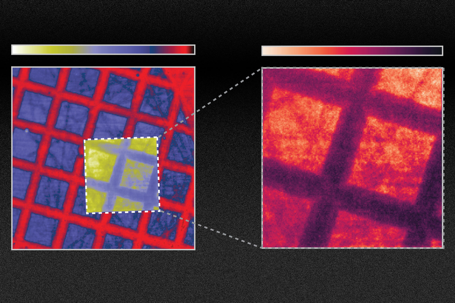
Researchers at MIT have now shown how one could improve the efficiency of scintillators by at least tenfold, and perhaps even a hundredfold, by changing the material’s surface to create certain nanoscale configurations, such as arrays of wave-like ridges. While past attempts to develop more efficient scintillators have focused on finding new materials, the new approach could in principle work with any of the existing materials.
Though it will require more time and effort to integrate their scintillators into existing X-ray machines, the team believes that this method might lead to improvements in medical diagnostic X-rays or CT scans, to reduce dose exposure and improve image quality. In other applications, such as X-ray inspection of manufactured parts for quality control, the new scintillators could enable inspections with higher accuracy or at faster speeds.
The findings are described today in the journal Science , in a paper by MIT doctoral students Charles Roques-Carmes and Nicholas Rivera; MIT professors Marin Soljacic, Steven Johnson, and John Joannopoulos; and 10 others.
While scintillators have been in use for some 70 years, much of the research in the field has focused on developing new materials that produce brighter or faster light emissions. The new approach instead applies advances in nanotechnology to existing materials. By creating patterns in scintillator materials at a length scale comparable to the wavelengths of the light being emitted, the team found that it was possible to dramatically change the material’s optical properties.
To make what they coined “nanophotonic scintillators,” Roques-Carmes says, “you can directly make patterns inside the scintillators, or you can glue on another material that would have holes on the nanoscale. The specifics depend on the exact structure and material.” For this research, the team took a scintillator and made holes spaced apart by roughly one optical wavelength, or about 500 nanometers (billionths of a meter).
“The key to what we’re doing is a general theory and framework we have developed,” Rivera says. This allows the researchers to calculate the scintillation levels that would be produced by any arbitrary configuration of nanophotonic structures. The scintillation process itself involves a series of steps, making it complicated to unravel. The framework the team developed involves integrating three different types of physics, Roques-Carmes says. Using this system they have found a good match between their predictions and the results of their subsequent experiments.
The experiments showed a tenfold improvement in emission from the treated scintillator. “So, this is something that might translate into applications for medical imaging, which are optical photon-starved, meaning the conversion of X-rays to optical light limits the image quality. [In medical imaging,] you do not want to irradiate your patients with too much of the X-rays, especially for routine screening, and especially for young patients as well,” Roques-Carmes says.
“We believe that this will open a new field of research in nanophotonics,” he adds. “You can use a lot of the existing work and research that has been done in the field of nanophotonics to improve significantly on existing materials that scintillate.”
“The research presented in this paper is hugely significant,” says Rajiv Gupta, chief of neuroradiology at Massachusetts General Hospital and an associate professor at Harvard Medical School, who was not associated with this work. “Nearly all detectors used in the $100 billion [medical X-ray] industry are indirect detectors,” which is the type of detector the new findings apply to, he says. “Everything that I use in my clinical practice today is based on this principle. This paper improves the efficiency of this process by 10 times. If this claim is even partially true, say the improvement is two times instead of 10 times, it would be transformative for the field!”
Soljacic says that while their experiments proved a tenfold improvement in emission could be achieved in particular systems, by further fine-tuning the design of the nanoscale patterning, “we also show that you can get up to 100 times [improvement] in certain scintillator systems, and we believe we also have a path toward making it even better,” he says.
Soljacic points out that in other areas of nanophotonics, a field that deals with how light interacts with materials that are structured at the nanometer scale, the development of computational simulations has enabled rapid, substantial improvements, for example in the development of solar cells and LEDs. The new models this team developed for scintillating materials could facilitate similar leaps in this technology, he says.
Nanophotonics techniques “give you the ultimate power of tailoring and enhancing the behavior of light,” Soljacic says. “But until now, this promise, this ability to do this with scintillation was unreachable because modeling the scintillation was very challenging. Now, this work for the first time opens up this field of scintillation, fully opens it, for the application of nanophotonics techniques.” More generally, the team believes that the combination of nanophotonic and scintillators might ultimately enable higher resolution, reduced X-ray dose, and energy-resolved X-ray imaging.
This work is “very original and excellent,” says Eli Yablonovitch, a professor of Electrical Engineering and Computer Sciences at the University of California at Berkeley, who was not associated with this research. “New scintillator concepts are very important in medical imaging and in basic research.”
Yablonovitch adds that while the concept still needs to be proven in a practical device, he says that, “After years of research on photonic crystals in optical communication and other fields, it's long overdue that photonic crystals should be applied to scintillators, which are of great practical importance yet have been overlooked” until this work.
The research team included Ali Ghorashi, Steven Kooi, Yi Yang, Zin Lin, Justin Beroz, Aviram Massuda, Jamison Sloan, and Nicolas Romeo at MIT; Yang Yu at Raith America, Inc.; and Ido Kaminer at Technion in Israel. The work was supported, in part, by the U.S. Army Research Office and the U.S. Army Research Laboratory through the Institute for Soldier Nanotechnologies, by the Air Force Office of Scientific Research, and by a Mathworks Engineering Fellowship.
Share this news article on:
Related links.
- Marin Soljacic
- Charles Roques-Carmes
- Nicholas Rivera
- John Joannopoulos
- Department of Physics
- Department of Mathematics
- Research Laboratory of Electronics
Related Topics
- Nanoscience and nanotechnology
- Artificial intelligence
- Electronics
- Materials science and engineering
- Health sciences and technology
Related Articles

Chip design drastically reduces energy needed to compute with light

Generating high-quality single photons for quantum computing

New system allows optical “deep learning”
Material could bring optical communication onto silicon chips
Previous item Next item
More MIT News

Study: Heavy snowfall and rain may contribute to some earthquakes
Read full story →

How AI might shape LGBTQIA+ advocacy

Two MIT PhD students awarded J-WAFS fellowships for their research on water

Exploring the mysterious alphabet of sperm whales

“Pathways to Invention” documentary debuts on PBS, streaming

This sound-suppressing silk can create quiet spaces
- More news on MIT News homepage →
Massachusetts Institute of Technology 77 Massachusetts Avenue, Cambridge, MA, USA
- Map (opens in new window)
- Events (opens in new window)
- People (opens in new window)
- Careers (opens in new window)
- Accessibility
- Social Media Hub
- MIT on Facebook
- MIT on YouTube
- MIT on Instagram
- Patient Care & Health Information
- Tests & Procedures
An X-ray is a quick, painless test that captures images of the structures inside the body — particularly the bones.
X-ray beams pass through the body. These beams are absorbed in different amounts depending on the density of the material they pass through. Dense materials, such as bone and metal, show up as white on X-rays. The air in the lungs shows up as black. Fat and muscle appear as shades of gray.
For some types of X-ray tests, a contrast medium — such as iodine or barium — is put into the body to get greater detail on the images.
Products & Services
- A Book: Mayo Clinic Family Health Book, 5th Edition
- Newsletter: Mayo Clinic Health Letter — Digital Edition
Why it's done

X-ray of knee arthritis
Knee arthritis can affect one side of the joint more than the other. This X-ray image shows how the cushioning cartilage has worn away, allowing bone to touch bone.
- Chest X-ray
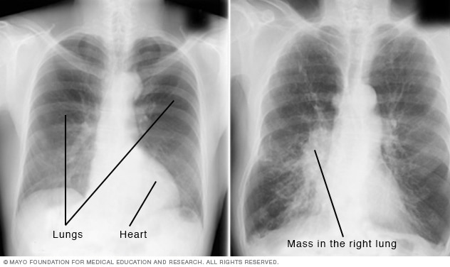
A chest X-ray helps detect problems with the heart and lungs. The chest X-ray on the left is typical. The image on the right shows a mass in the right lung.
- X-ray of swallowed jack
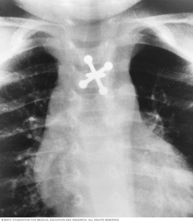
X-rays can locate metal objects your child has swallowed, such as this jack.
X-ray technology is used to examine many parts of the body.
Bones and teeth
- Fractures and infections. In most cases, fractures and infections in bones and teeth show up clearly on X-rays.
- Arthritis. X-rays of the joints can show evidence of arthritis. X-rays taken over the years can help your healthcare team tell if your arthritis is worsening.
- Dental decay. Dentists use X-rays to check for cavities in the teeth.
- Osteoporosis. Special types of X-ray tests can measure bone density.
- Bone cancer. X-rays can reveal bone tumors.
- Lung infections or conditions. Evidence of pneumonia, tuberculosis or lung cancer can show up on chest X-rays.
- Breast cancer. Mammography is a special type of X-ray test used to examine breast tissue.
- Enlarged heart. This sign of congestive heart failure shows up clearly on X-rays.
- Blocked blood vessels. Injecting a contrast material that contains iodine can help highlight sections of the circulatory system so they can be seen easily on X-rays.
- Digestive tract issues. Barium, a contrast medium delivered in a drink or an enema, can help show problems in the digestive system.
- Swallowed items. If a child has swallowed something such as a key or a coin, an X-ray can show the location of that object.
More Information
- Acanthosis nigricans
- Acute coronary syndrome
- Acute lymphocytic leukemia
- Adult Still disease
- Ambiguous genitalia
- Anal cancer
- Ankylosing spondylitis
- Anorexia nervosa
- Aspergillosis
- Atrial fibrillation
- Atrial septal defect (ASD)
- Avascular necrosis (osteonecrosis)
- Bed-wetting
- Bell's palsy
- Bladder stones
- Blastocystis hominis
- Bone cancer
- Breast cancer
- Broken ankle
- Broken collarbone
- Broken foot
- Broken hand
- Broken nose
- Broken ribs
- Broken wrist
- Brucellosis
- Bulimia nervosa
- Carcinoid tumors
- Carpal tunnel syndrome
- Castleman disease
- Cavities and tooth decay
- Cervical spondylosis
- Chagas disease
- Chronic exertional compartment syndrome
- Churg-Strauss syndrome
- Colon cancer
- Complex regional pain syndrome
- Constipation
- Constipation in children
- Craniopharyngioma
- Craniosynostosis
- Cystic fibrosis
- Dislocated shoulder
- Enlarged spleen (splenomegaly)
- Epiglottitis
- Esophageal cancer
- Esophageal spasms
- Frozen shoulder
- Functional neurologic disorder/conversion disorder
- Ganglion cyst
- Gastroesophageal reflux disease (GERD)
- Glomerulonephritis
- Golfer's elbow
- Growing pains
- Growth plate fractures
- Hamstring injury
- Herniated disk
- Hip dysplasia
- Hip fracture
- Hip labral tear
- Hirschsprung's disease
- Hodgkin's lymphoma (Hodgkin's disease)
- Horner syndrome
- Hyperparathyroidism
- Hypoparathyroidism
- Impacted wisdom teeth
- Indigestion
- Infant reflux
- Inflammatory bowel disease (IBD)
- Intestinal obstruction
- Intussusception
- Invasive lobular carcinoma
- Juvenile idiopathic arthritis
- Knee bursitis
- Legg-Calve-Perthes disease
- Lung cancer
- Male breast cancer
- Meralgia paresthetica
- Metatarsalgia
- Morton's neuroma
- Mouth cancer
- Multiple myeloma
- Neuroblastoma
- Neurofibromatosis
- Non-Hodgkin's lymphoma
- Osteoarthritis
- Osteochondritis dissecans
- Osteomyelitis
- Paget's disease of bone
- Paget's disease of the breast
- Patellar tendinitis
- Patellofemoral pain syndrome
- Peptic ulcer
- Peyronie disease
- Plantar fasciitis
- Posterior vaginal prolapse (rectocele)
- Precocious puberty
- Pseudomembranous colitis
- Psoriatic arthritis
- Pulmonary atresia
- Pulmonary atresia with intact ventricular septum
- Pulmonary atresia with ventricular septal defect
- Pyloric stenosis
- Reactive arthritis
- Recurrent breast cancer
- Residual limb pain
- Rotator cuff injury
- Sacroiliitis
- Sarcoidosis
- Septic arthritis
- Shaken baby syndrome
- Shin splints
- Spinal cord injury
- Spinal stenosis
- Sprained ankle
- Stress fractures
- Swollen knee
- Takayasu's arteritis
- Tapeworm infection
- Tennis elbow
- Thoracic outlet syndrome
- Throat cancer
- Thumb arthritis
- Tooth abscess
- Torn meniscus
- Ulcerative colitis
- Umbilical hernia
- Vaginal cancer
- Vascular dementia
- Vocal cord paralysis
Radiation exposure
Some people worry that X-rays aren't safe. This is because radiation exposure can cause cell changes that may lead to cancer. The amount of radiation you're exposed to during an X-ray depends on the tissue or organ being examined. Sensitivity to the radiation depends on your age, with children being more sensitive than adults.
Generally, however, radiation exposure from an X-ray is low, and the benefits from these tests far outweigh the risks.
However, if you are pregnant or suspect that you may be pregnant, tell your healthcare team before having an X-ray. Though most diagnostic X-rays pose only small risk to an unborn baby, your care team may decide to use another imaging test, such as ultrasound.
Contrast medium
In some people, the injection of a contrast medium can cause side effects such as:
- A feeling of warmth or flushing.
- A metallic taste.
- Lightheadedness.
Rarely, severe reactions to a contrast medium occur, including:
- Very low blood pressure.
- Difficulty breathing.
- Swelling of the throat or other parts of the body.
How you prepare
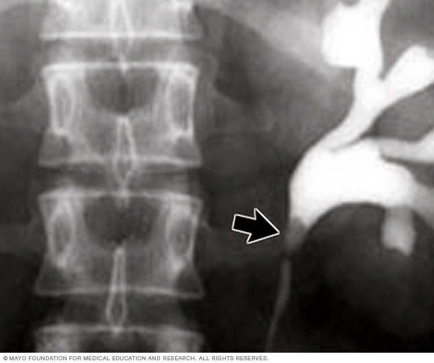
- X-ray image of kidney stone
This X-ray using contrast reveals a kidney stone at the junction of the kidney and the tube that connects the kidney to the bladder, called the ureter.
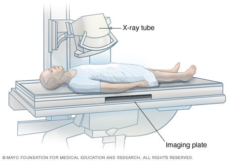
The X-ray tube is focused on the abdomen. X-rays will pass through the body and produce an image on the specialized plate below.
Different types of X-rays require different preparations. Ask your healthcare team to provide you with specific instructions.
What to wear
In general, you undress whatever part of your body needs examination. You may wear a gown during the exam depending on which area is being X-rayed. You also may be asked to remove jewelry, eyeglasses and any metal objects because they can show up on an X-ray.
Contrast material
Before having some types of X-rays, you're given a liquid called contrast medium. Contrast mediums, such as barium and iodine, help outline a specific area of your body on the X-ray image. You may swallow the contrast medium or receive it as an injection or an enema.
What you can expect
During the x-ray.
X-rays are performed at medical offices, dentists' offices, emergency rooms and hospitals — wherever an X-ray machine is available. The machine produces a safe level of radiation that passes through the body and records an image on a specialized plate. You can't feel an X-ray.
A technologist positions your body to get the necessary views. Pillows or sandbags may be used to help you hold the position. During the X-ray exposure, you remain still and sometimes hold your breath to avoid moving so that the image doesn't blur.
An X-ray procedure may take just a few minutes for a simple X-ray or longer for more-involved procedures, such as those using a contrast medium.
Your child's X-ray
If a young child is having an X-ray, restraints or other tools may be used to keep the child still. These won't harm the child and they prevent the need for a repeat procedure, which may be necessary if the child moves during the X-ray exposure.
You may be allowed to remain with your child during the test. If you remain in the room during the X-ray exposure, you'll likely be asked to wear a lead apron to shield you from unnecessary X-ray exposure.
After the X-ray
After an X-ray, you generally can resume usual activities. Routine X-rays usually have no side effects. However, if you're given contrast medium before your X-ray, drink plenty of fluids to help rid your body of the contrast. Call your healthcare team if you have pain, swelling or redness at the injection site. Ask your team about other symptoms to watch for.
X-rays are saved digitally on computers and can be viewed on-screen within minutes. A radiologist typically views and interprets the results and sends a report to a member of your healthcare team, who then explains the results to you. In an emergency, your X-ray results can be made available in minutes.
- X-rays. National Institute of Biomedical Imaging and Bioengineering. https://www.nibib.nih.gov/science-education/science-topics/x-rays. Accessed Nov. 7, 2023.
- Patient safety: Contrast materials. Radiological Society of North America. https://www.radiologyinfo.org/en/info/safety-contrast. Accessed Nov. 7, 2023.
- Chest X-ray. Radiological Society of North America. https://www.radiologyinfo.org/en/info/chestrad. Accessed Nov. 7, 2023.
- Bone X-ray (radiography). Radiological Society of North America. https://www.radiologyinfo.org/en/info/bonerad. Accessed Nov. 7, 2023.
- Pediatric X-ray. Radiological Society of North America. https://www.radiologyinfo.org/en/info/pediatric-xray. Accessed Nov. 7, 2023.
- Panoramic dental X-ray. Radiological Society of North America. https://www.radiologyinfo.org/en/info/panoramic-xray. Accessed Nov. 7, 2023.
- Lee CI, et al. Radiation-related risks of imaging. https://www.uptodate.com/contents/search. Accessed Nov. 7, 2023.
- Bone metastasis
- Cervical cancer
- Chronic cough
- Chronic pelvic pain
- Knee arthritis
- Monoclonal gammopathy of undetermined significance (MGUS)
- Nasopharyngeal carcinoma
- Peritonitis
- Rheumatoid arthritis
- Soft tissue sarcoma
- Spontaneous coronary artery dissection (SCAD)
- TMJ disorders
- X-ray during pregnancy
- Doctors & Departments
Mayo Clinic does not endorse companies or products. Advertising revenue supports our not-for-profit mission.
- Opportunities
Mayo Clinic Press
Check out these best-sellers and special offers on books and newsletters from Mayo Clinic Press .
- Mayo Clinic on Incontinence - Mayo Clinic Press Mayo Clinic on Incontinence
- The Essential Diabetes Book - Mayo Clinic Press The Essential Diabetes Book
- Mayo Clinic on Hearing and Balance - Mayo Clinic Press Mayo Clinic on Hearing and Balance
- FREE Mayo Clinic Diet Assessment - Mayo Clinic Press FREE Mayo Clinic Diet Assessment
- Mayo Clinic Health Letter - FREE book - Mayo Clinic Press Mayo Clinic Health Letter - FREE book
Your gift holds great power – donate today!
Make your tax-deductible gift and be a part of the cutting-edge research and care that's changing medicine.

X-ray astronomy is a relatively new science.
It was pioneered in the 1960s and 70s by American astronomers with a series of rocket flights and orbiting X-ray satellites. The astounding discoveries made by X-ray astronomers — such as neutron stars and black holes in binary systems, and hot gas filling the space within clusters of galaxies — have revolutionized our view of the Universe.
On July 23, 1999, NASA’s Chandra X-ray Observatory , the most powerful X-ray telescope ever built, was launched into space. Since then, Chandra has made numerous amazing discoveries, giving us a view of the Universe that is largely hidden from view through telescopes that observe in other types of light.
The technology developed for X-ray astronomy has evolved at a rapid pace, and has, moreover, produced numerous spinoff applications used in security monitoring, medicine and bio-medical research, materials processing, semi-conductor and microchip manufacturing, and environmental monitoring.

X-ray technology is now used in a wide variety of applications and settings. These include:
This area makes extensive use of X-ray technology spinoffs. The two major developments influenced by X-ray astronomy are the use of sensitive detectors to provide low dose but high-resolution images, and the linkage with digitizing and image processing systems. Many diagnostic procedures, such as mammographies and osteoporosis scans, require multiple exposures. It is important that each dosage be as low as possible. Accurate diagnosis also depends on the ability to view the subject from many different angles. Image processing systems linked to detectors capable of recording single X-ray photons, like those developed for X-ray astronomy purposes, provide doctors with the required data manipulation and enhancement capabilities. Smaller hand-held imaging systems can be used in clinics and under field conditions to diagnose sports injuries, to conduct outpatient surgery, and in the care of premature and new-born babies.
Biomedical Research
X-ray diffraction is the technique where X-ray light changes its direction by amounts that depend on the X-ray energy, much like a prism separates light into its component colors. Scientists using Chandra take advantage of diffraction to reveal important information about distant cosmic sources using the observatory’s two gratings instruments, the High Energy Transmission Grating Spectrometer (HETGS) and the Low Energy Transmission Grating Spectrometer (LETGS). X-ray diffraction is also used in biomedical and pharmaceutical research to study complex molecular structures. In most applications, the subject molecule is crystallized and then irradiated. The resulting diffraction pattern establishes the composition of the material. X-rays are perfect for this work because of their ability to resolve small objects. Advances in detector sensitivity and focused beam optics have allowed for the development of systems where exposure times have been shortened from hours to seconds. Shorter exposures coupled with lower-intensity radiation have allowed researchers to prepare smaller crystals, avoid damage to samples, and speed up their data runs. These systems are used for basics research with viruses, proteins, vaccines, and drugs, as well as for cancer, AIDS, and immunology research.
X-ray microscopy is a developing application. The microscope is, in effect, a miniature X-ray telescope. These microscopes have very high spatial resolution over small fields of view and they can be used to directly image very small images and fine details. Their applications are to energy and biomedical research.
Low Current Magnets
One of the instruments developed for use on Chandra was an X-ray spectrometer that would precisely measure the energy signatures over a key range of X-rays. In order to make these observations, this X-ray spectrometer had to be cooled to extremely low temperatures. Researchers at the Goddard Space Flight Center developed an innovative magnet that could achieve these very cold temperatures using a fraction of the helium that other similar magnets needed, thus extending the lifetime of the instrument’s use in space. On Earth, these advancements had benefits for MRI systems, making them safer and allowing for less maintenance.

Airport security
The first X-ray baggage inspection system for airports used detectors nearly identical to those flown in the Apollo program to measure fluorescent X-rays from the Moon. Its design took advantage of the sensitivity of the detectors that enabled the size, power requirements, and radiation exposure of the system to be reduced to limits practical for public use, while still providing adequate resolution to effectively screen baggage. The same company later developed a system that can simultaneously image, on two separate screens, materials of high atomic weight (e.g. metal hand guns) and materials of low atomic weight (e.g. plastic explosives) that pass through other systems undetected. Variations on these machines are used to screen visitors to public buildings around the world.

Quality Control and Manufacturing
Combining advanced X-ray detectors with image displays has resulted in a number of systems that can be used to inspect the quality of goods being produced or packaged on a production line. With these systems, the goods do not have to be brought to a special screening area and the production line does not have to be disrupted. The systems range from portable, hand-held models to large automated systems. They are used on such products as aircraft and rocket parts and structures, canned and packaged foods, electronics, semiconductors and microchips, thermal insulations, and automobile tires. Material processing technologies use X-ray systems to automatically control the density or thickness of layers of substances. Industries employing these systems include semiconductors and microchips, paper, tires, and oil and gas drilling.

Environmental Monitoring
Sensitive X-ray detectors coupled to automated data recording systems are used to monitor radon gas and radioactive waste. Low dose X-ray systems are also used to monitor tagged marine animals in environmental studies.

Energy Research
Researchers studying fusion as a potential source of energy use methods similar to those used by X-ray astronomers studying space plasmas. Very sensitive detector systems record the amount and location of the energy released by the fusion reaction in narrow slices of time. Theorists then interpret these observations to refine models for the fusion process and to advance the chances for obtaining significant amounts of usable energy from controlled fusion.
Lithography
X-ray beam lithography can produce extremely fine lines. Several companies are researching the use of focused X-ray synchrotron beam as the energy source for this process, since these powerful beams produce good pattern definition with relatively short exposure times. The grazing incidence optics developed for Chandra were the highest precision X-ray optics in the world and directly influenced this work.

Structure and Dynamics of Matter
Neutron scattering is one of the most useful methods for studying the structure and dynamics of matter. Lacking electrical charge neutrons penetrate deep inside materials. Neutron-scattering measurements can probe the atomic coordinates in crystal lattices, and the molecular conformation of polymers. Neutrons are especially sensitive to light elements such as hydrogen. Consequently, neutron radiography is used to measure water distribution in roots of growing plants or in very thin (~10 microns) membranes enclosed inside working fuel cells. Inspired by mirrors developed for X-ray astronomy, scientists have developed a new generation of neutron focusing optics for these and other applications.
For more information on applications from X-ray Astronomy development, please visit the web site for NASA's Technology Transfer Program .
Chandra blog: what chandra & x-ray astronomy give back.
- The Science of X-ray Technology - The Science of X-ray Technology Bookmark - The Science of X-ray Technology 6X6
Shareables:

If you have information on additional ways X-ray astronomy technology has been developed into other uses, we'd like to hear about it! Please email us at [email protected] .

SLAC researchers take important step toward developing cavity-based X-ray laser technology
They used diamond mirrors to make X-ray pulses run laps inside a vacuum chamber, demonstrating a key process needed for future generations of performance-enhanced X-ray lasers.
By Glennda Chui
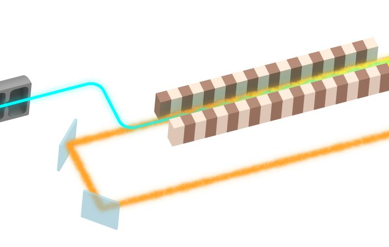
Researchers have announced an important step in the development of a next-gen technology for making X-ray free-electron laser pulses brighter and more stable: They used precisely aligned mirrors made of high-quality synthetic diamond to steer X-ray laser pulses around a rectangular racetrack inside a vacuum chamber.
Setups like these are at the heart of cavity-based X-ray free-electron lasers, or CBXFELs, which scientists are designing to make X-ray laser pulses brighter and cleaner – more like regular laser beams are today.
“The successful delivery of a cavity-based X-ray free-electron laser will mark the start of a new generation of X-ray science by providing a huge leap in beam performance,” said Mike Dunne, director of the Linac Coherent Light Source ( LCLS ) X-ray laser at the Department of Energy’s SLAC National Accelerator Laboratory, where the work was carried out.
“There are still many challenges to overcome before we get there,” he said. “But demonstration of this first integrated step is very encouraging, showing that we have the approach and tools needed to sustain high cavity performance.”
The SLAC research team described their work in a paper published in Nature Photonics. Early results were so encouraging, they said, that the lab is already working with DOE’s Argonne National Laboratory, its longtime collaborator on the subject, to design and install the next, bigger version of the experimental cavity system in the LCLS undulator tunnel.
Making X-ray laser pulses more laser-like
Despite their name, X-ray laser pulses are not yet fully laser-like. They’re created by making accelerated electrons wiggle through sets of magnets called undulators. This forces them to give off X-rays, which are shaped into powerful pulses for probing matter at the atomic scale. At LCLS, pulses arrive 120 times a second, a rate that will soon increase to a million times per second.
But because of the way X-ray laser pulses are generated, they vary in intensity and contain an unpredictable mix of wavelengths. This creates what scientists call “noise,” which muddles their view of samples they are probing.
The introduction of a cavity has been proposed to overcome this problem, adopting the approach used by conventional optical lasers. Cavities increase the coherence of lasers by preferentially selecting light of a single wavelength whose peaks and troughs line up with each other. But the mirrors that bounce light around in regular laser cavities won’t work for X-ray laser pulses – all you would get would be a smoking hole in your mirror where the X-rays drilled through.
The idea of using crystals – and, more recently, synthetic diamond crystals – as mirrors to smooth and help amplify X-ray pulses inside a cavity has been around for a long time, said Diling Zhu, who led the experimental team with fellow SLAC scientist Gabriel Marcus.
“The question was how to produce diamond mirrors of high enough quality and how to line them up with enough precision to steer the X-rays around the cavity,” Zhu said. “Ideally, in our case, the cavity would also fit into the long, narrow tunnel that houses the LCLS undulators.”
Additional challenges and innovations include finding the best way to take X-rays out of the cavity so they can be used for experiments and to optimally cool the mirrors, if needed.
A barbell-like setup
The SLAC cavity project started about five years ago with a few hallway conversations, Zhu said. Those led to a Laboratory Directed Research and Development grant from the SLAC director to build the setup used in this study in an LCLS experimental hutch.
“What’s unique about this experiment is the large scale at which it was done. It’s nearly 50 times bigger than any other version I’ve seen published,” said Rachel Margraf, a Stanford graduate student and one of the researchers who co-designed and carried out the experiment and analyzed the results.
“The bigger the cavity is,” she said, “the tighter the alignment tolerances are, and the scientific community had been skeptical that those tolerances could be achieved.”
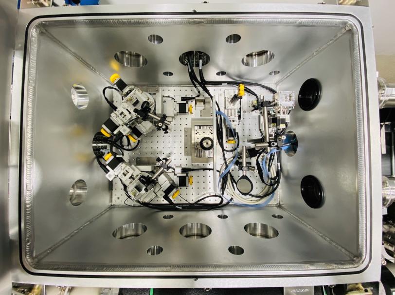
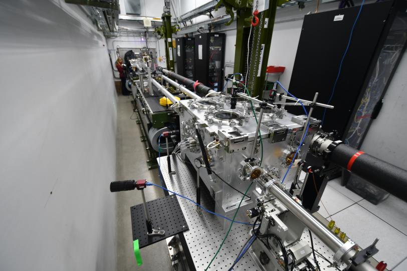
The experimental setup consists of two boxy vacuum chambers that contain the cavity components. They’re connected by two beam pipes, which are also kept under vacuum. From the side, the whole thing resembles a 30-foot-long barbell.
Each cavity chamber houses two diamond mirrors, and each mirror is mounted on a set of four motors that precisely adjust its position and angle to the beam. The mirrors steer X-ray pulses through the beam pipes and from one mirror to the next.
Crafting the perfect diamond mirror
Synthesizing, selecting and shaping the diamond mirrors was a big effort in itself.
The diamonds were prepared by Kenji Tamasaku, team leader of the XFEL division at the RIKEN SPring-8 Center in Japan and a world authority on diamonds for X-ray research, in collaboration with an industry partner.
Growing diamond crystals that are pure enough for X-ray research is tricky, Tamasaku said, because they have to be grown at high temperatures and pressures where the slightest change in conditions can disrupt the crystal growth.
The team first used X-ray microscopes from SPring-8 and the Stanford Synchrotron Radiation Light Source (SSRL) at SLAC to carefully examine each crystal and cherry-pick the ones with the fewest defects in their crystal structure. Then they identified areas within those crystals that were defect-free for processing into mirrors.
“The quality of natural diamonds cannot compete with that of diamonds used in the present study,” Tamasaku said.
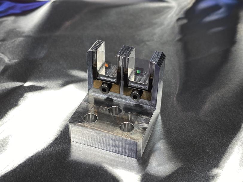
Near-perfect bits of diamond crystal were cut with lasers, first into slabs and then into S shapes about one-fifth of an inch long that were polished to a high shine, a process first developed by experts at Argonne. The mirrors have tags that can be clamped onto the experimental apparatus without putting strain on the mirror itself.
Successful results
The purpose of this experiment was to see how long and how efficiently X-ray laser pulses could circulate inside the cavity. LCLS laser pulses entered the setup 120 times per second via a precision diamond grating crafted on the Stanford campus. They hit each of the mirrors in turn, completing up to 60 laps – each about 46 feet long – before dissipating. The researchers said the round-trip travel efficiency within the cavity setup was more than 96% – close to the theoretical mirror performance limit, and more than sufficient to support a high-quality X-ray laser beam.
Ultimately, the goal is to store and circulate X-ray pulses in the cavity and then send them through the undulator to accompany the electron beam as it wiggles through the magnets. Repeating this cycle 10 to 100 times should create X-ray laser beams that are as coherent and stable as today’s optical laser beams, Zhu said. This will become possible with the completion of LCLS upgrades that substantially increase the energy and repetition rate of its X-ray laser pulses. “We are super excited about working with our Argonne and RIKEN colleagues to get another step closer to this goal,” Zhu said.
LCLS and SSRL are DOE Office of Science user facilities, and funding for the construction and installation of the cavity system prototype at LCLS comes from the Office of Science. The diamond grating that lets X-ray pulses into the cavity was crafted on the Stanford campus by the SLAC Nano-X team.
Citation : Rachel Mar graf et al., Nature Photonics , 14 August 2023 ( 10.1038/s41566-023-01267-0 )
For questions or comments, contact the SLAC Office of Communications at [email protected] .
Related Topics
- Science news
- Advanced accelerator R and D
- Ultrafast science
- X-ray science
- X-ray light sources and electron imaging
- Future light sources
- Linac Coherent Light Source (LCLS)
- Stanford Synchrotron Radiation Lightsource (SSRL)
Related stories
Recommendations for u.s. government investments in particle physics include slac research priorities.
A new report outlines suggestions for federal investments needed for the next generation of transformative discoveries in particle physics and cosmology, including priority projects...
Shrinking particle accelerators with cold plasma and a large picnic basket
What could smaller particle accelerators look like in the future? SLAC scientists are working on innovations that could give more researchers access to accelerator...
Physicists ask: Can we make a particle collider more energy efficient?
The future of experimental particle physics is exciting – and energy intensive. SLAC physicists are thinking about how to make one proposal, the Cool...
SLAC fires up the world’s most powerful X-ray laser: LCLS-II ushers in a new era of science
With up to a million X-ray flashes per second, 8,000 times more than its predecessor, it transforms the ability of scientists to explore atomic-scale...

Researchers show how to increase X-ray laser brightness and power using a crystal cavity and diamond mirrors
The long – but not too long – cavity would ping-pong X-ray pulses inside of a particle accelerator facility to help capture nature’s fastest...
Researchers develop clever algorithm to improve our understanding of particle beams in accelerators
The algorithm pairs machine-learning techniques with classical beam physics equations to avoid massive data crunching.


X-RAYS AND ENERGY
X-rays have much higher energy and much shorter wavelengths than ultraviolet light, and scientists usually refer to x-rays in terms of their energy rather than their wavelength. This is partially because x-rays have very small wavelengths, between 0.03 and 3 nanometers, so small that some x-rays are no bigger than a single atom of many elements.

DISCOVERY OF X-RAYS
X-rays were first observed and documented in 1895 by German scientist Wilhelm Conrad Roentgen. He discovered that firing streams of x-rays through arms and hands created detailed images of the bones inside. When you get an x-ray taken, x-ray sensitive film is put on one side of your body, and x-rays are shot through you. Because bones are dense and absorb more x-rays than skin does, shadows of the bones are left on the x-ray film while the skin appears transparent.
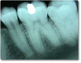
An x-ray image of teeth. Can you see the filling?
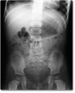
An X-ray photo of a one year old girl who swallowed a sewing pin. Can you find it?
Our Sun's radiation peaks in the visual range, but the Sun's corona is much hotter and radiates mostly x-rays. To study the corona, scientists use data collected by x-ray detectors on satellites in orbit around the Earth. Japan's Hinode spacecraft produced these x-ray images of the Sun that allow scientists to see and record the energy flows within the corona.
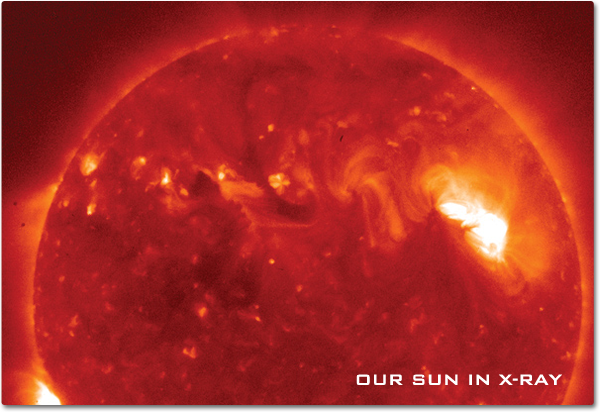
TEMPERATURE AND COMPOSITION
The physical temperature of an object determines the wavelength of the radiation it emits. The hotter the object, the shorter the wavelength of peak emission. X-rays come from objects that are millions of degrees Celsius—such as pulsars, galactic supernovae remnants, and the accretion disk of black holes.
From space, x-ray telescopes collect photons from a given region of the sky. The photons are directed onto the detector where they are absorbed, and the energy, time, and direction of individual photons are recorded. Such measurements can provide clues about the composition, temperature, and density of distant celestial environments. Due to the high energy and penetrating nature of x-rays, x-rays would not be reflected if they hit the mirror head on (much the same way that bullets slam into a wall). X-ray telescopes focus x-rays onto a detector using grazing incidence mirrors (just as bullets ricochet when they hit a wall at a grazing angle).
NASA's Mars Exploration Rover, Spirit, used x-rays to detect the spectral signatures of zinc and nickel in Martian rocks. The Alpha Proton X-Ray Spectrometer (APXS) instrument uses two techniques, one to determine structure and another to determine composition. Both of these techniques work best for heavier elements such as metals.

Since Earth's atmosphere blocks x-ray radiation, telescopes with x-ray detectors must be positioned above Earth's absorbing atmosphere. The supernova remnant Cassiopeia A (Cas A) was imaged by three of NASA's great observatories, and data from all three observatories were used to create the image shown below. Infrared data from the Spitzer Space Telescope are colored red, optical data from the Hubble Space Telescope are yellow, and x-ray data from the Chandra X-ray Observatory are green and blue.
The x-ray data reveal hot gases at about ten million degrees Celsius that were created when ejected material from the supernova smashed into surrounding gas and dust at speeds of about ten million miles per hour. By comparing infrared and x-ray images, astronomers are learning more about how relatively cool dust grains can coexist within the super-hot, x-ray producing gas.

EARTH'S AURORA IN X-RAYS
Solar storms on the Sun eject clouds of energetic particles toward Earth. These high-energy particles can be swept up by Earth's magnetosphere, creating geomagnetic storms that sometimes result in an aurora. The energetic charged particles from the Sun that cause an aurora also energize electrons in the Earth's magnetosphere. These electrons move along the Earth's magnetic field and eventually strike the Earth's ionosphere, causing the x-ray emissions. These x-rays are not dangerous to people on the Earth because they are absorbed by lower parts of the Earth's atmosphere. Below is an image of an x-ray aurora by the Polar Ionospheric X-ray Imaging Experiment (PIXIE) instrument aboard the Polar satellite.
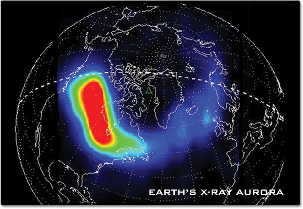
Next: Gamma Rays
National Aeronautics and Space Administration, Science Mission Directorate. (2010). X-Rays. Retrieved [insert date - e.g. August 10, 2016] , from NASA Science website: http://science.nasa.gov/ems/11_xrays
Science Mission Directorate. "X-Rays" NASA Science . 2010. National Aeronautics and Space Administration. [insert date - e.g. 10 Aug. 2016] http://science.nasa.gov/ems/11_xrays
Discover More Topics From NASA
James Webb Space Telescope

Perseverance Rover

Parker Solar Probe

An official website of the United States government
The .gov means it's official. Federal government websites often end in .gov or .mil. Before sharing sensitive information, make sure you're on a federal government site.
The site is secure. The https:// ensures that you are connecting to the official website and that any information you provide is encrypted and transmitted securely.
- Publications
- Account settings
- Browse Titles
NCBI Bookshelf. A service of the National Library of Medicine, National Institutes of Health.
StatPearls [Internet]. Treasure Island (FL): StatPearls Publishing; 2024 Jan-.

StatPearls [Internet].
X-ray production.
Dawood Tafti ; Christopher V. Maani .
Affiliations
Last Update: July 31, 2023 .
- Definition/Introduction
X-rays are a form of electromagnetic radiation with wavelengths ranging from 0.01 to 10 nanometers. In the setting of diagnostic radiology, X-rays have long enjoyed use in the imaging of body tissues and aid in the diagnosis of disease. Simply understood, the generation of X-rays occurs when electrons are accelerated under a potential difference and turned into electromagnetic radiation. [1] An X-ray tube, with its respective components placed in a vacuum, and a generator, make up the basic components of X-ray production. Essential components of an X-ray tube include a cathode, and an anode separated a short distance from each other, a vacuum enclosure, and high voltage cables forming the X-ray generator attached to the cathode and anode components. [2] In the generation of X-ray production, a cathode filament machined in a cathode cup is activated, causing intense heating of the cathode filament. [3] The heating of the filament leads to the release of electrons in a process called thermionic emission. [4] The released electrons form in an electron cloud at the filament surface, and repulsion forces prevent the ejection of electrons from this negatively charged cloud. [2] Upon application of a high voltage by an X-ray generator to the cathode as well as the anode, there is an acceleration of electrons ejected to an electrically positive anode. [3] The filament and the focusing cup determine this path of acceleration. The number of electrons is measured in the form of milliampere (mA) units, where 1 milliampere is equal to 6.24 x 10^15 electrons/s. Electron kinetic energy (measured in keV) is related to the applied voltage. The tube voltage, tube current, and exposure duration (measured in seconds) are adjustable by the user.
Once the high kinetic energy electrons finally reach the anode target, this initiates the process of X-ray production. Tungsten is often the usual anode target, although other material targets are also employed. Electrons come extremely close to the nucleus of the target, causing a deceleration and change in direction, converting the kinetic energy to electromagnetic radiation in a process known as “breaking radiation” or bremsstrahlung. [5] The output is a spectrum of X-ray energies. Incident electrons can also result in ionization, whereby the approaching electron can remove a second electron belonging to an atom of the anode target, losing its energy through ionization or excitation. This process leads to an emission of a photon as the electron orbit vacancy gets filled by an orbital shell electron from a further out shell. Considering orbital energies and their differences are unique in atoms, this leads to a “characteristic X-ray” with energies that can serve as a fingerprint unique to each anode target. Bremsstrahlung X-rays, however, constitute the majority of X-rays produced in this process.
Before understanding the final production of an X-ray image, it is essential to understand the interaction of X-rays with individuals exposed to X-rays. There are three important types of interactions that occur between X-rays and the tissues of our body. The “classical” or “coherent” interaction occurs when an X-ray strikes an orbital electron and subsequently bounces off and changes direction. [6] These X-rays are low energy and do not cause ionization and only add a small dose amount to a patient. In “Compton” scattering, X-rays of higher energy strike an outer shell electron and are strong enough to remove it from the shell, causing ionization of an atom. [7] This phenomenon contributes to dose and also contributes to scatter. Photoelectric interactions occur when an incoming X-ray strikes an inner shell electron, removing it from the shell and causing a downward cascade of outer shell electrons filling inner orbit vacancies, further releasing secondary X-rays. This type of interaction contributes to image contrast. Finally, the differential absorption of X-rays within the tissues of the body subsequently contributes to the production of the final image. Attenuation of X-rays ultimately depends on the effective atomic number in tissue, X-ray beam energy, and tissue density. [8]
Image detectors come in the form of digital and analog film detectors. [9] One limitation of analog systems is the limited range of exposure intensities that it can accurately detect; this lends itself to multiple images taken for an adequate and interpretable study, and therefore subsequently leads to increased radiation exposure to a patient. Digital systems allow a user to fix contrast and brightness and provide greater post image processing options. [9]
- Issues of Concern
Effective dose refers to the amount of radiation received by the whole body, and measurement is in millisievert (mSv). Generally speaking, various procedures entail different effective radiation doses based on site and use of contrast. For example, a radiograph of the spine has an approximate effective dose of around 1 mSv. [10] A radiograph of the extremity ranges within the upper limits of normal between 0.17 to 2.7 microSv. [10] To better place these doses in context, we can compare these exposures with natural radiation we obtain from our surroundings, which usually approximates to 3 mSv per year. [11] A spine X-ray, therefore, is comparable to the natural background radiation exposure for six months. An extremity radiograph compares to natural background radiation exposure of 3 hours. Bone densitometry and mammography studies have an approximate effective dose of around 0.001 mSv and 0.4 mSv, respectively, comparable to 3 hours and six weeks of background radiation, respectively. [12] Radiography, therefore, in the setting of cumulative exposure is not without risks in patients who require frequent imaging studies. An X-ray technician plays an instrumental role in the acquisition of interpretable and high-quality X-ray images. A working and constant relationship between a radiologist and an X-ray technician is essential in troubleshooting and acquiring images in the appropriate diagnosis of a patient. X-ray technicians are also critical in preventing artifacts, taking brief medical histories, ensuring appropriate laterality, appropriate positioning, and adjusting and maintaining various equipment involved in X-ray acquisition.
- Clinical Significance
Although adequate coverage of the full range of uses of conventional radiographs cannot is beyond the scope of this article, the use of radiography frequently plays a critical role in assessing the various osseous structures of the body. Evaluation of the lungs is also possible, and the use of contrast can also help to examine soft tissue organs of the body, including the gastrointestinal tract and the uterus, such as in the setting of hysterosalpingography. Radiography is useful in performing various procedures including catheter angiography, stereotactic breast biopsies as well as an intra-articular steroid injection. Radiography helps in the evaluation of multiple pathologies, including fractures, types of pneumonia, malignancies, as well as congenital anatomic abnormalities.
- Review Questions
- Access free multiple choice questions on this topic.
- Comment on this article.
X-ray generator. Contributed by Dawood Tafti, MD
Disclosure: Dawood Tafti declares no relevant financial relationships with ineligible companies.
Disclosure: Christopher Maani declares no relevant financial relationships with ineligible companies.
This book is distributed under the terms of the Creative Commons Attribution-NonCommercial-NoDerivatives 4.0 International (CC BY-NC-ND 4.0) ( http://creativecommons.org/licenses/by-nc-nd/4.0/ ), which permits others to distribute the work, provided that the article is not altered or used commercially. You are not required to obtain permission to distribute this article, provided that you credit the author and journal.
- Cite this Page Tafti D, Maani CV. X-ray Production. [Updated 2023 Jul 31]. In: StatPearls [Internet]. Treasure Island (FL): StatPearls Publishing; 2024 Jan-.
In this Page
Bulk download.
- Bulk download StatPearls data from FTP
Related information
- PMC PubMed Central citations
- PubMed Links to PubMed
Similar articles in PubMed
- Electrostatic focal spot correction for x-ray tubes operating in strong magnetic fields. [Med Phys. 2014] Electrostatic focal spot correction for x-ray tubes operating in strong magnetic fields. Lillaney P, Shin M, Hinshaw W, Fahrig R. Med Phys. 2014 Nov; 41(11):112302.
- Radiology, Image Production and Evaluation. [StatPearls. 2024] Radiology, Image Production and Evaluation. Nakashima J, Duong H. StatPearls. 2024 Jan
- The AAPM/RSNA physics tutorial for residents. X-ray production. [Radiographics. 1997] The AAPM/RSNA physics tutorial for residents. X-ray production. McCollough CH. Radiographics. 1997 Jul-Aug; 17(4):967-84.
- Study of increased radiation when an x-ray tube is placed in a strong magnetic field. [Med Phys. 2007] Study of increased radiation when an x-ray tube is placed in a strong magnetic field. Wen Z, Pelc NJ, Nelson WR, Fahrig R. Med Phys. 2007 Feb; 34(2):408-18.
- Review X-ray imaging physics for nuclear medicine technologists. Part 1: Basic principles of x-ray production. [J Nucl Med Technol. 2004] Review X-ray imaging physics for nuclear medicine technologists. Part 1: Basic principles of x-ray production. Seibert JA. J Nucl Med Technol. 2004 Sep; 32(3):139-47.
Recent Activity
- X-ray Production - StatPearls X-ray Production - StatPearls
Your browsing activity is empty.
Activity recording is turned off.
Turn recording back on
Connect with NLM
National Library of Medicine 8600 Rockville Pike Bethesda, MD 20894
Web Policies FOIA HHS Vulnerability Disclosure
Help Accessibility Careers
- Type 2 Diabetes
- Heart Disease
- Digestive Health
- Multiple Sclerosis
- Diet & Nutrition
- Supplements
- Health Insurance
- Public Health
- Patient Rights
- Caregivers & Loved Ones
- End of Life Concerns
- Health News
- Thyroid Test Analyzer
- Doctor Discussion Guides
- Hemoglobin A1c Test Analyzer
- Lipid Test Analyzer
- Complete Blood Count (CBC) Analyzer
- What to Buy
- Editorial Process
- Meet Our Medical Expert Board
What Is an X-Ray?
What to Expect When Undergoing This Test
How It Works
- When It's Used
Contraindications
How to prepare, during the test, after the test, interpreting results.
An X-ray, also known as radiography , is a medical imaging technique. It uses tiny amounts of electromagnetic radiation to create images of structures inside the body. These images can then be viewed on film or digitally.
X-rays often are done to view bones and teeth , making them helpful in diagnosing fractures (broken bones) and diseases such as arthritis . A healthcare provider may also order an X-ray to look at organs and structures inside the chest, including the lungs, heart, breasts, and abdomen.
This article explains when X-rays are used, how to prepare for one, and what to expect. It also covers the risks and benefits of the imaging test.
The tiny particles of electromagnetic radiation that an X-ray machine emits pass through all but the most solid objects in the body. As such, the image it creates, known as a radiograph, allows healthcare providers to visualize internal structures in your body.
What Is Electromagnetic Radiation?
Electromagnetic radiation (EMR) is a type of radiation that travels in waves and has electric and magnetic fields. Devices that use this type of radiation include X-rays, microwaves, radio waves, ultraviolet light, infrared light, visible light, and gamma rays.
Sometimes a contrast medium , a type of dye, is given to help images appear in greater detail. You might receive these via injection into a blood vessel, orally, or rectally.
X-ray images appear in various shades of white and grey. Because bones and metal objects are solid, less radiation passes through them, making them appear white on the radiograph. On the other hand, skin, muscle, blood and other fluids, and fat are grey because they allow most radiation to pass through.
Areas where there is nothing to stop the beam of radiation, such as air, or even a fracture, appear black compared to surrounding tissue.
When It's Used
X-ray technology is used for a multitude of purposes. For example, it can help healthcare providers evaluate symptoms and diagnose injuries.
Among the most common reasons for X-rays include:
- Identifying fractures
- Identifying infections in bones and teeth
- Diagnosing cavities and evaluating structures in the mouth and jaw
- Revealing bone tumors
- Measuring bone density (the amount of mineral in your bones) to diagnose osteoporosis (a bone disease caused by bone loss)
- Finding evidence of pneumonia, tuberculosis , or lung cancer
- Looking for signs of heart failure or changes in blood flow to the lungs and heart
- Revealing problems in the digestive tract, such as kidney stones , sometimes using a contrast medium called barium
- Locating swallowed items such as a coin or tiny toy
This technology can also support other types of diagnostic procedures.
Fluoroscopy
During fluoroscopy , an X-ray image displays on a monitor in real time. Unlike X-ray images, which are still pictures, fluoroscopy is a moving image. Often, you will receive a contrast dye intravenously (in your vein) during this procedure.
Seeing moving images allows healthcare providers to follow the progression of a procedure (such as the placement of a stent). They can also view the contrast agent passing through the body.
Computed tomography (CT scan) is a technique that takes a series of individual images of internal organs and tissues. These “slices” are then combined to show a three-dimensional visualization.
CT scans can identify organ masses, see how well blood is flowing, observe brain hemorrhage and trauma, view lung structures, and diagnose injuries and diseases of the skeletal system.
Mammography
A mammogram is a breast cancer screening test that uses X-ray imaging. Mammograms can also diagnose breast lumps and other breast changes.
During a mammogram, your breasts are placed one at a time between two plates. A technician then presses them together to flatten your breast to get a clear picture. Finally, they X-ray your breasts from the front and sides.
Arthrography allows healthcare providers to identify signs of joint changes that indicate arthritis. It uses an X-ray and a special contrast dye injected directly into the joint.
Sometimes instead of X-rays, an arthrogram uses CT scan, fluoroscopy, or magnetic resonance imaging (MRI) technology.
Having an X-ray doesn't hurt and isn't particularly dangerous. However, there are a few things to be aware of and discuss with your healthcare provider.
Radiation Exposure
Having frequent X-rays carries a very low risk of developing cancer later in life. That is because the radiation has enough energy to potentially damage DNA .
There are varying estimates as to how significant this risk is. What is known is that fluoroscopy and computed tomography both expose the body to more radiation than a single conventional X-ray. The Food and Drug Administration (FDA) says that the risk of cancer from exposure to X-rays depends on:
- Exposure frequency
- Age at exposure
- Which reproductive organs a person has
- Area of the body exposed
The more times a person is exposed to radiation from medical imaging throughout their life and the larger the dose, the greater the risk of developing cancer. In addition, the lifetime risk of cancer is more significant for someone who's exposed to radiation at a younger age than for a person who has X-rays when they're older.
Studies have shown that those with female reproductive organs are at a somewhat higher lifetime risk for developing radiation-associated cancer. Researchers believe that since reproductive organs absorb more radiation and people with ovaries typically have more reproductive organs than those with testicles, this may be why.
It is essential to weigh the risks and benefits of having an X-ray, CT scan, or fluoroscopy with your healthcare provider. Ask if the imaging study will make an impact on your care. If not, it may be advisable to skip the test. However, if a diagnosis or potential changes in your treatment are likely to depend on the X-ray results, it will most likely be worth the minor risk.
Barium Sulfate Risks
There may be some minor risks associated with contrast mediums used during X-ray procedures, particularly for people who have asthma or other conditions.
Barium sulfate contrast materials are perfectly safe for most people. However, some circumstances can put a person at an increased risk of severe side effects such as throat swelling, difficulty breathing, and more. These include:
- Having asthma or allergies , which increases the risk of an allergic reaction
- Cystic fibrosis , which increases the risk of small bowel blockage
- Severe dehydration, which may cause severe constipation
- An intestinal blockage or perforation that could be made worse by the contrast agent
Iodine Risks
Iodine is another contrast medium used for X-rays. After exposure to this dye, a small percentage of people may develop delayed reaction hours or even days later. Most reactions are mild, but some can be more severe and cause the following:
- Skin rash or hives
- Abnormal heart rhythms
- High or low blood pressure
- Shortness of breath
- Difficulty breathing
- Throat swelling
- Cardiac arrest
- Convulsions
Given your overall health profile, a healthcare provider can help you determine if using a contrast agent is necessary and suitable for you.
Pregnant people are usually discouraged from having an X-ray unless it's vital. That's because there is a risk that the radiation from an X-ray could cause changes in developing fetal cells and thereby increase the risk of birth defects or cancer later in life. The risk of harm depends on a fetus's gestational age and the amount of radiation exposure.
That said, this recommendation is mainly precautionary. These risks are associated with very high doses of radiation, and a regular diagnostic X-ray does not expose you to high-dose radiation. Therefore, the benefits of what an X-ray could reveal often outweigh any risks.
If you need an X-ray during pregnancy, the following can reduce your risks:
- Cover with a leaded apron or collar to block any scattered radiation
- Avoid abdominal X-rays
- Inform the X-ray technician if you are or could be pregnant
In addition, if you have a child who needs an X-ray, don't hold them during the procedure if you are or might be pregnant.
Often, an X-ray is done as part of a visit to a healthcare provider or emergency room to diagnose symptoms or evaluate an injury. X-rays also complement specific routine exams, such as dental checkups . These types of X-rays usually take place in a medical office or the hospital.
Other times, a healthcare provider recommends screening X-rays, like mammograms, at regular intervals. These are often performed at imaging centers or hospitals by appointment.
The setting in which you get an X-ray and its reasons will determine your overall testing experience.
It's impossible to generalize how long an entire X-ray procedure will take. For example, it can take just a few minutes to get an image or two of an injured bone in an emergency room. On the other hand, a CT scan appointment can take longer.
If you're scheduling an X-ray, ask your healthcare provider to give you an idea of how much time you should allow.
X-ray tests may take place in various locations, including:
- Hospital imaging departments
- Freestanding radiology and imaging clinics
- Medical offices, particularly specialists such as orthopedics and dentists
- Urgent care centers
What to Wear
Generally speaking, the X-ray tech will ask you to remove any clothing covering the X-rayed area. For some procedures that involve X-ray imaging, you'll need to wear a hospital gown. Therefore, you may want to choose clothing that's easy to change in and out of.
In addition, since metal can show up on an X-ray, you may need to remove your jewelry and eyeglasses before an X-ray.
Food and Drink
If you have an X-ray without contrast, you can usually eat and drink normally. However, if you are receiving a contrast agent, you may need to avoid consuming food and liquids for some time before.
For example, healthcare providers use barium to highlight structures in the digestive system . Therefore, they may tell you not to eat for at least three hours before your appointment.
People with diabetes are usually advised to eat a light meal three hours before receiving barium. However, suppose you receive the barium via an enema (a tube inserted into the rectum). In that case, you may also be asked to eat a special diet and take medication to cleanse your colon beforehand.
Cost and Health Insurance
Most health insurance policies will cover any medically necessary X-ray imaging. Of course, out-of-pocket costs vary and depend on the type of plan you have. For example, you may be responsible for a copay , or for the entire cost if you haven't met your deductible . Check with your insurance company to learn the specifics of your plan.
If you don't have insurance or you're paying out-of-pocket for an X-ray, the fee will depend on several things, including:
- Which body part is imaged
- The number of images taken
- Whether a contrast dye is used
Similarly, if you are paying for your X-ray and have time to research the fees, you can call the hospital's billing department ahead of time to get a quote for the procedure. Doing so can help you know the cost you're obligated to pay.
What to Bring
You will need to have your insurance card with you at your X-ray. In addition, if your healthcare provider gave you a written order for the procedure, bring that as well.
Because X-ray procedures vary widely, it isn't easy to generalize the experience. So instead, ask your healthcare provider for details about what to expect in your specific case.
You may need to remove some or all of your clothing before the X-ray. A technician will escort you to a dressing room or other private area where you can change into a hospital gown. There will probably be a locker where you can safely store your clothing and other belongings.
If you have a test involving a contrast dye, you will receive that before your imaging procedure.
Healthcare providers may give contrast dyes in the following ways:
- In a special drink you swallow
- Intravenous (IV) line
Except for IV contrast dye, which allows for a constant stream of the material to be given, contrasts are administered before the X-ray. In other words, you will not have to wait for the dye to "take" before your imaging test.
How you receive the contrast depends on the substance used and what internal organs or structures your healthcare provider needs to view. For example, you might receive an iodine-based contrast dye injection into a joint for an arthrogram.
On the other hand, you might swallow a barium contrast to help illuminate your digestive system for fluoroscopy. Oral barium contrast dye may not taste good, but most people can tolerate the flavor long enough to swallow the prescribed amount.
If you have a barium enema, you may feel abdominal fullness and urgency to expel the liquid. However, the mild discomfort will not last long.
A conventional X-ray is taken in a special room with an X-ray machine. During the test, you will:
- Place a leaded apron or cover over your torso
- Stand, sit, or lie down on an X-ray table
- Position your body in specific ways
- Use props such as sandbags or pillows to adjust your position
Once positioned correctly, you will need to be very still. That's because even slight movement can cause an X-ray image to come out blurry. A technician may even ask you to hold your breath.
Infants and young children may need support being still. Guardians often accompany small children into the procedure room for this reason. If you attend your child for support, you will wear a leaded apron to limit your radiation exposure.
For their protection, the technician will step behind a protective window to operate the X-ray machine while also watching you. It only takes a few seconds to take the picture. However, often multiple angles of the body part are necessary. So, after your first image, the technician will likely adjust you or the machine and take another picture.
For a CT scan, you will lie down on a table that moved you into a cylindrical machine that rotates around you to take many pictures from all directions . You won't feel anything during a CT scan, but it may be uncomfortable for you if you dislike being in enclosed spaces.
When the tech has all the required images, you will remove the lead apron (if used) and leave the room. If you need to change back into your clothes, they'll direct you to the dressing area to change out of your hospital gown.
After you leave your appointment, you can return to your regular activities. If you received a contrast medium, a healthcare provider might instruct you to drink extra fluids to help flush the substance out of your system.
The barium-based dye comes out in your bowel movements, which will be white for a few days. You also may notice changes in your bowel movement patterns for 12 to 24 hours after your X-ray.
If you take Glucophage ( metformin ) or a related medication to treat type 2 diabetes, you need to stop taking your medicine for at least 48 hours after receiving contrast. That's because it may cause a condition called metabolic acidosis—an unsafe change in your blood pH (the balance of acidic or alkaline substances in the body).
Barium Side Effects
Keep an eye on the injection site if you received contrast dye by injection. Call your healthcare provider if you experience signs of infection, like pain, swelling, or redness.
Barium contrast materials can cause some digestive tract problems. If these become severe or don't go away, see your healthcare provider. These side effects include:
- Stomach cramps
- Nausea and vomiting
- Constipation
Iodine Side Effects
Likewise, iodine contrast can cause symptoms. Let your healthcare provider know if you begin to have even mild symptoms after receiving iodine contrast. These symptoms include:
- Mild skin rash and hives
Severe Side Effects
Call your healthcare provider immediately or go to the emergency room if you experience signs of anaphylaxis , a severe allergic reaction, including:
- Swelling of the throat
- Difficulty breathing or swallowing
- Fast heartbeat
- Bluish skin color
A radiologist specializing in analyzing imaging tests interprets the images from your X-ray. They then send the results and a report to your healthcare provider. Often, they will call you or have you come in to discuss the findings. In emergencies, you should receive these results soon after your X-ray.
Any follow-up tests or treatment will depend on your particular situation. For example, if you have an X-ray to determine the extent of an injury to a bone and it reveals you have a break, the bone will need to be set . Likewise, a breast tumor revealed during mammography may require a follow-up biopsy to determine if it is malignant (cancerous) or benign (non-cancerous).
X-rays are imaging tests that use small amounts of electromagnetic radiation to view the inside structures of your body. In addition to conventional X-rays, several other specialized forms of X-rays capture images in more precise ways. Sometimes a contrast agent can help healthcare providers see things better. These dyes might be given via injection, IV, orally, or rectally.
X-rays don't typically require preparation unless you are receiving contrast. In that case, you may need to avoid food and drinks for a few hours beforehand. X-rays do not take long—usually just a few minutes. Often, a technician takes multiple angles and images of the area. Afterward, you will be able to leave right away. If you received contrast, you might notice side effects. You should tell your healthcare provider about any symptoms you experience.
A Word From Verywell
For the majority of people, X-rays are harmless. However, if you have to have multiple X-rays over a lifetime, you may be at increased cancer risk. As such, it's essential to talk to your healthcare provider before you have an X-ray to make sure you have all the information you need to make an informed decision. And if you are or could be pregnant, tell the technician before undergoing the procedure.
National Cancer Institute. Electromagnetic radiation .
U.S. National Library of Medicine MedlinePlus. X-rays .
U.S. Food & Drug Administration. Medical x-ray imaging .
Johns Hopkins Medicine. Arthrography .
Narendran N, Luzhna L, Kovalchuk O. Sex difference of radiation response in occupational and accidental exposure . Front Genet . 2019;10:260. doi:10.3389/fgene.2019.00260
RadiologyInfo.org. Contrast materials .
U.S. Food & Drug Administration. X-rays, pregnancy and you .
RadiologyInfo.org. X-ray (radiography) .
RadiologyInfo.org. Pediatric X-ray (radiography) .
Johns Hopkins Medicine. What are X-rays?
U.S. Food and Drug Administration. Medical X-ray imaging .
By Kristin Hayes, RN Kristin Hayes, RN, is a registered nurse specializing in ear, nose, and throat disorders for both adults and children.
For the best browsing experience please enable JavaScript. Instructions for Microsoft Edge and Internet Explorer , other browsers

- About cancer
- Get involved
- Our research
- Funding for researchers
- Cancer types
- Cancer in general
- Causes of cancer
- Coping with cancer
- Health professionals
- Do your own fundraising
- By cancer type
- By cancer subject
- Our funding schemes
- Applying for funding
- Managing your research grant
- How we deliver our research
- Find a shop
- Shop online
- Our eBay shop
- Our organisation
- Current jobs
- Cancer news

An x-ray is a test that uses small amounts (doses) of radiation to take pictures of the inside of your body. They are a good way to look at bones and can show changes caused by cancer or other medical conditions. X-rays can also show changes in other organs, such as the lungs.
You usually have x-rays in the imaging department of the hospital, taken by a radiographer. But in an emergency they are sometimes done on the ward.
There are different types of tests using x-rays, including:
- chest x-rays to show fluid, signs of infection, an enlarged heart or tumours in the chest such as lung cancer
- x-rays of the bones to show breaks (fractures), degenerative changes, infection or tumours
- x-rays of the breasts (mammograms)
- dental x-rays to look at the teeth and jaw
- real time x-ray screening (fluoroscopy) to help doctors put in stents or wires, or to look at blood vessels (angiography), or to show the outline of body structures (barium x-rays)
- CT scans are a series of x-rays of an area of the body to build up a 3 dimensional (3D) picture
What happens
There is no special preparation for a standard x-ray. You can eat and drink normally beforehand. You take your medicines as normal. If you are having another type of x-ray such as:
- a barium x-ray
- an angiogram
You might need to stop eating and drinking for a certain amount of time before the test. Your appointment letter will give you instructions you need to follow.
When you arrive, the radiographer might ask you to change into a hospital gown and take off any jewellery.
During your x-ray
You usually have a chest x-ray standing up against the x-ray machine. If you can’t stand you can have it sitting or lying on the x-ray couch. For x-rays of other areas of the body the best position is usually lying down on the x-ray couch.
The radiographer lines the machine up to make sure it's in the right place. You must keep very still to prevent blurring of the picture.
The radiographer then goes behind a screen. They can still see and hear you. They might ask you to hold your breath for a few seconds while they take the x-ray.
X-rays are painless and quick. You won’t feel anything.
You might have more than one x-ray taken from different angles. The whole process may take a few minutes.
After your x-ray
After the x-ray you can get dressed and go home or back to work.
Possible risks
An x-ray is a safe test for most people but like all medical tests it has some possible risks. Your doctor and radiographer make sure the benefits of having the test outweigh these risks.
The amount of radiation you receive from an x-ray is small and doesn't make you feel unwell.
The risk of the radiation causing any problems in the future is very small. The benefits of finding out what is wrong outweigh any risk there may be from radiation.
Talk to your doctor if you are worried about the possible effects of x-ray.
Fertility and pregnancy
The ovaries and testicles are particularly sensitive to radiation. You may have lead blocks to shield them if they are in the x-ray field.
It is very important to tell your doctor if you think you may be pregnant. X-rays could affect your developing baby. If you can’t delay the x-ray, the radiographer may be able to shield the baby with a lead apron or block.
Getting your results
You should get your results within 1 or 2 weeks at a follow up appointment.
Waiting for test results can be a very worrying time. You might have contact details for a specialist nurse who you can contact for information if you need to. It can help to talk to a close friend or relative about how you feel.
Contact the doctor who arranged the test if you haven’t heard anything after a couple of weeks.
More information
We have more information on tests, treatment and support if you have been diagnosed with cancer.
- Find information on your cancer type
Related links
Your cancer type.
Search for the cancer type you want to find out about. Each section has detailed information about symptoms, diagnosis, treatment, research and coping with cancer.
Tests and scans
Find out about tests to diagnose cancer and monitor it during and after treatment, including what each test can show, how you have it and how to prepare.
Barium x -ray
A barium x-ray is a test to look at the outline of any part of your digestive system. Find out how you have it and what happens afterwards.
A mammogram is an x-ray of your breasts. This test is used to help diagnose breast cancer and other breast conditions.
A CT scan is a test that uses x-rays and a computer to create detailed pictures of the inside of your body. Find out how you have it and what happens afterwards.
It’s a worrying time for many people and we want to be there for you whenever - and wherever - you need us. Cancer Chat is our fully moderated forum where you can talk to others affected by cancer, share experiences, and get support. Cancer Chat is free to join and available 24 hours a day.
Visit the Cancer Chat forum
About Cancer generously supported by Dangoor Education since 2010.

Find a clinical trial
Search our clinical trials database for all cancer trials and studies recruiting in the UK

Cancer Chat forum
Talk to other people affected by cancer
Nurse helpline 0808 800 4040
Questions about cancer? Call freephone 9 to 5 Monday to Friday or email us
share this!
April 29, 2024
This article has been reviewed according to Science X's editorial process and policies . Editors have highlighted the following attributes while ensuring the content's credibility:
fact-checked
trusted source
Einstein probe opens its wide eyes to the X-ray sky
by European Space Agency

The first images captured by the innovative mission were presented at the 7th workshop of the Einstein Probe consortium in Beijing. They illustrate the satellite's full potential and show that its novel optics, which mimic a lobster's eyes, are ready to monitor the X-ray sky. The space X-ray telescope zoomed in on a few well-known celestial objects to give us a hint of what the mission is capable of.
Launched on 9 January 2024, the Chinese Academy of Sciences (CAS) spacecraft Einstein Probe joins ESA's XMM-Newton and JAXA's XRISM in their quest to discover the universe in X-ray light. The mission is a collaboration led by CAS with ESA, the Max Planck Institute for Extraterrestrial Physics (MPE) (Germany), and the National Center for Space Studies (CNES) (France).
In the months since liftoff, the mission operations team has been performing the necessary tests to confirm the spacecraft's functionality and calibrating the scientific instruments. During this crucial phase, Einstein Probe captured scientific data from various X-ray sources.
These first-light images demonstrate the outstanding capabilities of Einstein Probe's two scientific instruments . The Wide-field X-ray Telescope (WXT) can observe a panorama of nearly one-eleventh of the celestial sphere in one shot, while the more sensitive Follow-up X-ray Telescope (FXT) offers close-ups and can pinpoint short-lived events caught by WXT.
"I am delighted to see the first observations from Einstein Probe, which showcase the mission's ability to study wide expanses of the X-ray sky and quickly discover new celestial sources," says Prof. Carole Mundell, ESA Director of Science.
"These early data give us a tantalizing glimpse of the high-energy dynamic universe that will soon be within reach of our science communities. Congratulations to the science and engineering teams at CAS, MPE, CNES and ESA for their hard work in reaching this important milestone."
The capability of the mission to promptly spot new X-ray sources and monitor how they change over time is fundamental to improving our grasp of the most energetic processes in the cosmos. Powerful X-rays are blasted through the universe when neutron stars collide, supernovas explode, and matter is swallowed by black holes or ejected from the crushing magnetic fields that envelop them.
Lobster eyes monitoring the universe
Einstein Probe's WXT instrument consists of 12 modules featuring the novel lobster-eye technology that was tested in flight in 2022 by the technology demonstrator LEIA (Lobster Eye Imager for Astronomy). The 12 modules provide a field of view of more than 3,600 square degrees, allowing Einstein Probe to monitor the whole night sky in just three orbits.

During its first months in space, WXT started its work of keeping a watchful eye on the X-ray sky. Detections of energetic objects look like a lit-up plus sign due to the way the instrument's novel lobster-eye optics work. The first X-ray transient source—an astronomical object that is not continuously shining but pops up and fades again—was discovered on 19 February. This candidate gamma-ray burst lasted for 100 seconds. Einstein Probe discovered another 14 temporary X-ray sources and also captured X-rays 127 flaring stars.
During the mission, the wide-field instrument's findings will guide a range of ground- and space-based telescopes to perform follow-up observations in multiple wavelength bands. X-ray follow-up observations can also be obtained using the satellite's FXT instrument.
Rapid follow-up observations
Einstein Probe's FXT instrument has a set of two X-ray telescopes for detailed studies of X-ray-emitting objects and events. During the past months, FXT has proved to be a trustworthy instrument to observe a range of X-ray sources. The first images bring into new focus a supernova remnant, an elliptical galaxy, a globular cluster and a nebula.
Remarkably, FXT already performed a follow-up observation of an X-ray event spotted by WXT on 20 March 2024.
"It is astounding that even though the instruments were not yet fully calibrated, we could already perform a time-critical follow-up observation using the FXT instrument of a fast X-ray transient first spotted by WXT," explains Dr. Erik Kuulkers, ESA's Einstein Probe Project Scientist. "It shows what Einstein Probe will be capable of during its survey."
What's next?
In the coming months, Einstein Probe will continue to undergo in-orbit calibration activities before starting its routine science observations around mid-June. During the three-year mission, the satellite will circle Earth at a height of 600 km and keep its eyes on the sky searching for transitory X-ray events. Using the FXT follow-up telescope, the mission will look deeper at newly detected events and other known interesting objects.
Einstein Probe's capabilities are highly complementary to the in-depth studies of individual cosmic sources enabled by XMM-Newton and XRISM. Its survey is fundamental to prepare for X-ray observations by ESA's future NewAthena mission, currently under study and set to be the largest X-ray observatory ever built.
Provided by European Space Agency
Explore further
Feedback to editors

Every drop counts: New algorithm tracks Texas's daily reservoir evaporation rates
2 hours ago

Genetic study finds early summer fishing can have an evolutionary impact, resulting in smaller salmon
5 hours ago

Researchers discovery family of natural compounds that selectively kill parasites

Study suggests heavy snowfall and rain may contribute to some earthquakes

The spread of misinformation varies by topic and by country in Europe, study finds

Webb presents best evidence to date for rocky exoplanet atmosphere

Human activity is making it harder for scientists to interpret oceans' past
6 hours ago

Quantum simulators solve physics puzzles with colored dots

Chemists produce new-to-nature enzyme containing boron

Improving timing precision of millisecond pulsars using polarization
Relevant physicsforums posts, dark matter and its effect on the orbit of stars.
53 minutes ago
Solar Activity and Space Weather Update thread
Why do novas shine.
23 hours ago
NASA names asteroid "Hossi" after Sabine Hossenfelder
May 7, 2024
Documenting the setup of my new telescope
Our beautiful universe - photos and videos.
May 6, 2024
More from Astronomy and Astrophysics
Related Stories

Einstein Probe lifts off on a mission to monitor the X-ray sky
Jan 9, 2024

Innovative X-ray lobster-eye mission set to launch
Dec 21, 2023

EP-WXT pathfinder catches first wide-field snapshots of X-ray universe
Sep 2, 2022

Another example of a fantastic Einstein ring
Jan 11, 2024

Next major X-ray mission set to launch
Sep 4, 2023

Image: First light data for NASA's Parker Solar Probe
Sep 20, 2018
Recommended for you

Astronomers explore globular cluster NGC 2419
14 hours ago

New accreting millisecond X-ray pulsar discovered

How NASA's Roman mission will hunt for primordial black holes

Radio astronomers bypass disturbing Earth's atmosphere with new calibration technique
Let us know if there is a problem with our content.
Use this form if you have come across a typo, inaccuracy or would like to send an edit request for the content on this page. For general inquiries, please use our contact form . For general feedback, use the public comments section below (please adhere to guidelines ).
Please select the most appropriate category to facilitate processing of your request
Thank you for taking time to provide your feedback to the editors.
Your feedback is important to us. However, we do not guarantee individual replies due to the high volume of messages.
E-mail the story
Your email address is used only to let the recipient know who sent the email. Neither your address nor the recipient's address will be used for any other purpose. The information you enter will appear in your e-mail message and is not retained by Phys.org in any form.
Newsletter sign up
Get weekly and/or daily updates delivered to your inbox. You can unsubscribe at any time and we'll never share your details to third parties.
More information Privacy policy
Donate and enjoy an ad-free experience
We keep our content available to everyone. Consider supporting Science X's mission by getting a premium account.
E-mail newsletter

Suggested Searches
- Climate Change
- Expedition 64
- Mars perseverance
- SpaceX Crew-2
- International Space Station
- View All Topics A-Z
Humans in Space
Earth & climate, the solar system.
- The Universe
Aeronautics
Learning resources, news & events.
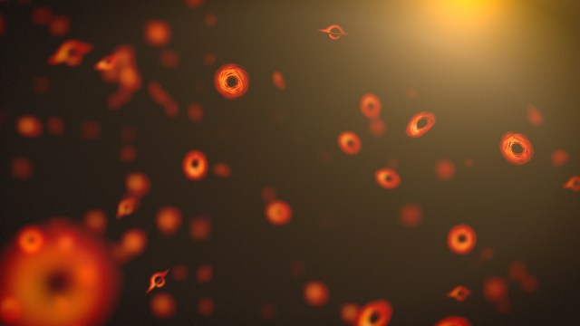
How NASA’s Roman Mission Will Hunt for Primordial Black Holes

International SWOT Mission Can Improve Flood Prediction
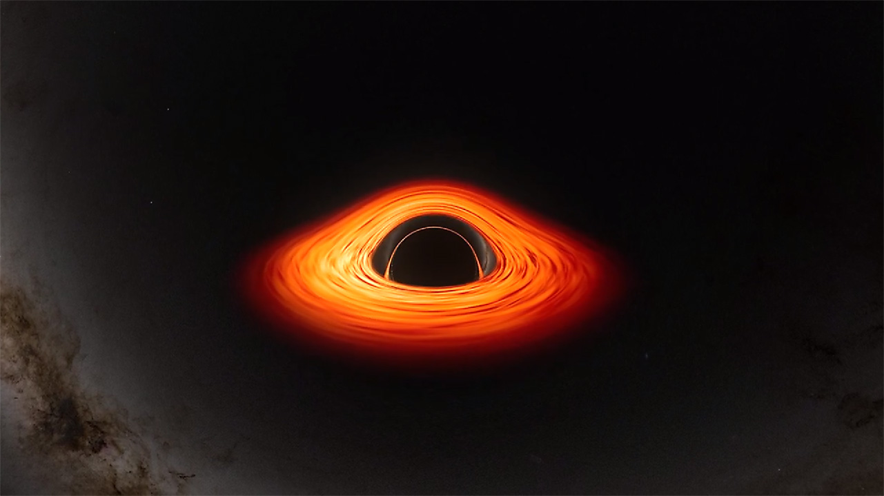
New NASA Black Hole Visualization Takes Viewers Beyond the Brink
- Search All NASA Missions
- A to Z List of Missions
- Upcoming Launches and Landings
- Spaceships and Rockets
- Communicating with Missions
- James Webb Space Telescope
- Hubble Space Telescope
- Why Go to Space
- Astronauts Home
- Commercial Space
- Destinations
- Living in Space
- Explore Earth Science
- Earth, Our Planet
- Earth Science in Action
- Earth Multimedia
- Earth Science Researchers
- Pluto & Dwarf Planets
- Asteroids, Comets & Meteors
- The Kuiper Belt
- The Oort Cloud
- Skywatching
- The Search for Life in the Universe
- Black Holes
- The Big Bang
- Dark Energy & Dark Matter
- Earth Science
- Planetary Science
- Astrophysics & Space Science
- The Sun & Heliophysics
- Biological & Physical Sciences
- Lunar Science
- Citizen Science
- Astromaterials
- Aeronautics Research
- Human Space Travel Research
- Science in the Air
- NASA Aircraft
- Flight Innovation
- Supersonic Flight
- Air Traffic Solutions
- Green Aviation Tech
- Drones & You
- Technology Transfer & Spinoffs
- Space Travel Technology
- Technology Living in Space
- Manufacturing and Materials
- Science Instruments
- For Kids and Students
- For Educators
- For Colleges and Universities
- For Professionals
- Science for Everyone
- Requests for Exhibits, Artifacts, or Speakers
- STEM Engagement at NASA
- NASA's Impacts
- Centers and Facilities
- Directorates
- Organizations
- People of NASA
- Internships
- Our History
- Doing Business with NASA
- Get Involved
- Aeronáutica
- Ciencias Terrestres
- Sistema Solar
- All NASA News
- Video Series on NASA+
- Newsletters
- Social Media
- Media Resources
- Upcoming Launches & Landings
- Virtual Events
- Sounds and Ringtones
- Interactives
- STEM Multimedia

NASA’s Webb Hints at Possible Atmosphere Surrounding Rocky Exoplanet

NASA, JAXA XRISM Spots Iron Fingerprints in Nearby Active Galaxy

International SWOT Mission Can Improve Flood Prediction

NASA Mission Strengthens 40-Year Friendship

NASA Selects Commercial Service Studies to Enable Mars Robotic Science

NASA’s Commercial Partners Deliver Cargo, Crew for Station Science

NASA Is Helping Protect Tigers, Jaguars, and Elephants. Here’s How.
C.26 Rapid Mission Design Studies for Mars Sample Return Correction and Other Documents Posted

NASA Selects Students for Europa Clipper Intern Program

Orbits and Kepler’s Laws

NASA’s TESS Returns to Science Operations

ARMD Solicitations

NASA’s Commitment to Safety Starts with its Culture
NASA Uses Small Engine to Enhance Sustainable Jet Research
Tech Today: NASA’s Ion Thruster Knowhow Keeps Satellites Flying
Big Science Drives Wallops’ Upgrades for NASA Suborbital Missions

NASA Challenge Gives Artemis Generation Coders a Chance to Shine
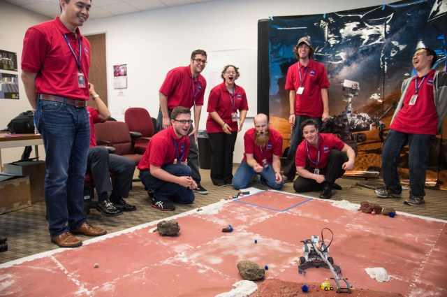
NASA Community College Aerospace Scholars

Ken Carpenter: Ensuring Top-Tier Science from Moon to Stars

White Sands Propulsion Team Tests 3D-Printed Orion Engine Component

A Different Perspective – Remembering James Dean, Founder of the NASA Art Program

Diez maneras en que los estudiantes pueden prepararse para ser astronautas

Astronauta de la NASA Marcos Berríos

Resultados científicos revolucionarios en la estación espacial de 2023
X-ray satellite xmm-newton sees ‘space clover’ in a new light.

Ashley Balzer
Astronomers have discovered enormous circular radio features of unknown origin around some galaxies. Now, new observations of one dubbed the Cloverleaf suggest it was created by clashing groups of galaxies.
Studying these structures, collectively called ORCs (odd radio circles), in a different kind of light offered scientists a chance to probe everything from supersonic shock waves to black hole behavior.
“This is the first time anyone has seen X-ray emission associated with an ORC,” said Esra Bulbul, an astrophysicist at the Max Planck Institute for Extraterrestrial Physics in Garching, Germany, who led the study. “It was the missing key to unlock the secret of the Cloverleaf’s formation.”
A paper describing the results was published in Astronomy and Astrophysics Letters on April 30.
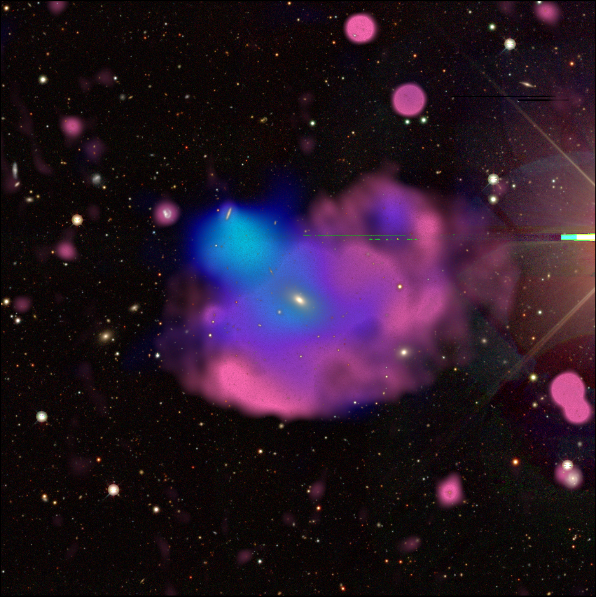
A Serendipitous Discovery
Until 2021 , no one knew ORCs existed. Thanks to improved technology, radio surveys became sensitive enough to pick up such faint signals. Over the course of a few years, astronomers discovered eight of these strange structures scattered randomly beyond our galaxy. Each is large enough to envelop an entire galaxy –– sometimes several.
“The power needed to produce such an expansive radio emission is very strong,” Bulbul said. “Some simulations can reproduce their shapes but not their intensity. No simulations explain how to create ORCs.”
When Bulbul learned ORCs hadn’t been studied in X-ray light, she and postdoctoral researcher Xiaoyuan Zhang began poring over data from eROSITA (Extended Roentgen Survey with an Imaging Telescope Array), an orbiting German/Russian X-ray telescope. They noticed some X-ray emission that seemed like it could be from the Cloverleaf, based on less than 7 minutes of observation time.
That gave them a strong enough case to assemble a larger team and secure additional telescope time with XMM-Newton, an ESA (European Space Agency) mission with NASA contributions.
“We were allotted about five-and-a-half hours, and the data came in late one evening in November,” Bulbul said. “I forwarded it to Xiaoyuan, and he came into my office the next morning and said, ‘Detection,’ and I just started cheering!”
“We really got lucky,” Zhang said. “We saw several plausible X-ray point sources close to the ORC in eROSITA observations, but not the expanded emission we saw with XMM-Newton. It turns out the eROSITA sources couldn’t have been from the Cloverleaf, but it was compelling enough to get us to take a closer look.”
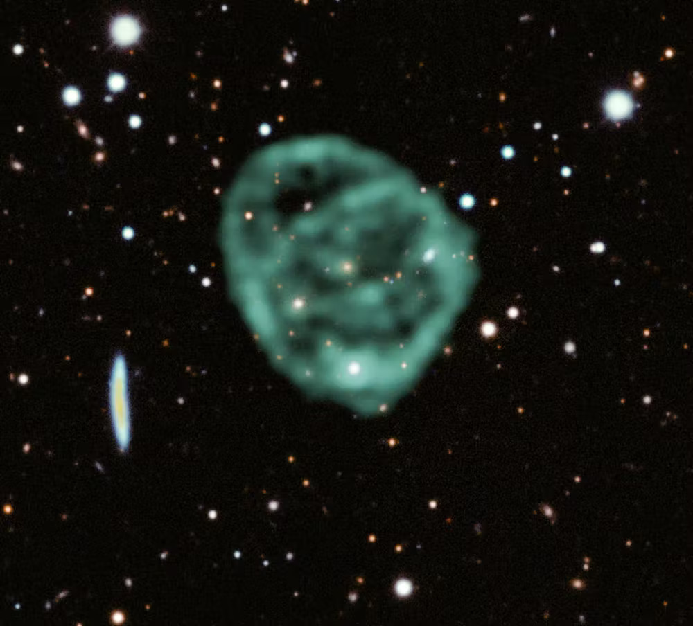
Gallivanting Galaxies
The X-ray emission traces the distribution of gas within the group of galaxies like police tape around a crime scene. By seeing how that gas has been disturbed, scientists determined that galaxies embedded in the Cloverleaf are actually members of two separate groups that drew close enough together to merge. The emission’s temperature also hints at the number of galaxies involved.
When galaxies join, their higher combined mass increases their gravity. Surrounding gas begins to fall inward, which heats up the infalling gas. The greater the system’s mass, the hotter the gas becomes.
Based on the emission’s X-ray spectrum, it’s around 15 million degrees Fahrenheit, or between 8 and 9 million degrees Celsius. “That measurement let us deduce that the Cloverleaf ORC is hosted by around a dozen galaxies that have gravitated together, which agrees with what we see in deep visible light images,” Zhang said.
The team proposes the merger produced shock waves that accelerated particles to create radio emission.
“Galaxies interact and coalesce all the time,” said Kim Weaver, the NASA project scientist for XMM-Newton at NASA’s Goddard Space Flight Center in Greenbelt, Maryland, who was not involved in the study. “But the source of the accelerated particles is unclear. One fascinating idea for the powerful radio signal is that the resident supermassive black holes went through episodes of extreme activity in the past, and relic electrons from that ancient activity were reaccelerated by this merging event.”
While galaxy group mergers are common, ORCs are very rare. And it’s still unclear how these interactions can produce such strong radio emissions.
“Mergers make up the backbone of structure formation, but there’s something special in this system that rockets the radio emission,” Bulbul said. “We can’t tell right now what it is, so we need more and deeper data from both radio and X-ray telescopes.”
The team solved the mystery of the nature of the Cloverleaf ORC, but also opened up additional questions. They plan to study the Cloverleaf in more detail to tease out answers.
“We stand to learn a lot from more thorough observations because these interactions take in all kinds of science,” Weaver says. “You’ve pretty much got everything that we deal with in the cosmos put together in this little package. It’s like a mini universe.”
For more information on ESA’s XMM-Newton mission, visit:
https://science.nasa.gov/mission/xmm-newton/
By Ashley Balzer NASA’s Goddard Space Flight Center , Greenbelt, Md.
Media Contact: Claire Andreoli 301-286-1940 [email protected] NASA’s Goddard Space Flight Center, Greenbelt, Md.
Explore More

X-ray Satellite XMM-Newton Celebrates 20 Years in Space

Swift XMM-Newton Satellites Tune Into a Middleweight Black Hole

Chandra and XMM-Newton Provide Direct Measurement of Distant Black Hole’s Spin
Related terms.
- XMM-Newton (X-ray Multi-Mirror Newton)
- Active Galaxies
- Astrophysics
- Elliptical Galaxies
- Galaxies, Stars, & Black Holes
- Galaxies, Stars, & Black Holes Research
- Galaxy clusters
- Goddard Space Flight Center
- Science & Research
- Sensing the Universe & Multimessenger Astronomy

A novel 3D high resolution imaging method using correlative X-Ray and electron microscopy to study Nd:YAG laser induced defects in intraocular lenses
- Split-Screen
- Article contents
- Figures & tables
- Supplementary Data
- Peer Review
- Open the PDF for in another window
- Get Permissions
- Cite Icon Cite
- Search Site
Andreas F. Borkenstein , Adrian Mikitisin , Alexander Schwedt , Eva-Maria Borkenstein , Joachim Mayer; A novel 3D high resolution imaging method using correlative X-Ray and electron microscopy to study Nd:YAG laser induced defects in intraocular lenses . Ophthalmic Res 2024; https://doi.org/10.1159/000539243
Download citation file:
- Ris (Zotero)
- Reference Manager
Article PDF first page preview
Introduction: Cataract extraction is the most frequently performed ophthalmological procedure worldwide. Posterior capsule opacification (PCO) remains the most common consequence after cataract surgery and can lead to deterioration of the visual performance with cloudy, blurred vision and halo, glare effects. Nd:YAG laser capsulotomy is the gold standard treatment and a very effective, safe and fast procedure in removing the cloudy posterior capsule. Damaging the intraocular lens during the treatment may occur due to wrong focus of the laser beam. These YAG-pits may lead to a permanent impairment of the visual quality. Methods: In an experimental study, we intentionally induced YAG pits in hydrophilic and hydrophobic acrylic intraocular lenses (IOLs) using a photodisruption laser with 2.6mJ. This experimental study established a novel 3D imaging method using correlative X-Ray and scanning electron microscopy to characterize these damages. By integrating the information obtained from both X-ray microscopy and SEM, a comprehensive picture of the materials structure and performance could be established. Results: It could be revealed that although the exact same energies were used to all samples, the observed defects in the tested lenses showed severe differences in shape and depth. While YAG pits in hydrophilic samples range from 100 to 180 µm depth with a round shape tip, very sharp tipped defects up to 250 µm in depth were found in hydrophobic samples. In all samples, particles/fragments of the IOL material were found on the surface that were blasted out as a result of the laser shelling. Conclusion: Defects in hydrophilic and hydrophobic acrylic materials differ. Material particles can detach from the IOL and were found on the surface of the samples. The results of the laboratory study illustrate the importance of a precise and careful approach to Nd:YAG capsulotomy in order to avoid permanent damage to the IOL. The use of an appropriate contact glass and posterior offset setting to increase safety should be carried out routinely.

Email alerts
Citing articles via.
- Online ISSN 1423-0259
- Print ISSN 0030-3747
INFORMATION
- Contact & Support
- Information & Downloads
- Rights & Permissions
- Terms & Conditions
- Catalogue & Pricing
- Policies & Information
- People & Organization
- Stay Up-to-Date
- Regional Offices
- Community Voice
SERVICES FOR
- Researchers
- Healthcare Professionals
- Patients & Supporters
- Health Sciences Industry
- Medical Societies
- Agents & Booksellers
Karger International
- S. Karger AG
- P.O Box, CH-4009 Basel (Switzerland)
- Allschwilerstrasse 10, CH-4055 Basel
- Tel: +41 61 306 11 11
- Fax: +41 61 306 12 34
- Contact: Front Office
- Experience Blog
- Privacy Policy
- Terms of Use
This Feature Is Available To Subscribers Only
Sign In or Create an Account
Thank you for visiting nature.com. You are using a browser version with limited support for CSS. To obtain the best experience, we recommend you use a more up to date browser (or turn off compatibility mode in Internet Explorer). In the meantime, to ensure continued support, we are displaying the site without styles and JavaScript.
- View all journals
Radiography articles from across Nature Portfolio
Radiography is the use of X-rays or gamma rays to examine non-uniformly composed material; images are recorded on a sensitized surface, such as photographic film or a digital detector. Medical radiography includes examination of any part of the body for diagnostic purposes, such as detecting fractures or a bowel obstruction.
Latest Research and Reviews
Quantitative analysis and stochastic modeling of osteophyte formation and growth process on human vertebrae based on radiographs: a follow-up study
- Changxi Wang
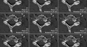
Influence of helical pitch and gantry rotation time on image quality and file size in ultrahigh-resolution photon-counting detector CT
- Philipp Feldle
- Jan-Peter Grunz
- Nora Conrads
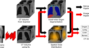
Two-view topogram-based anatomy-guided CT reconstruction for prospective risk minimization
- Laura Klein
- Andreas Maier
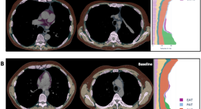
Body composition impacts outcome of bronchoscopic lung volume reduction in patients with severe emphysema: a fully automated CT-based analysis
- Johannes Wienker
- Kaid Darwiche
- Marcel Opitz
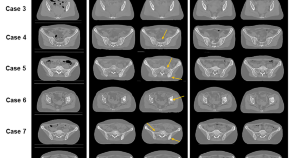
Clinical feasibility of deep learning-based synthetic CT images from T2-weighted MR images for cervical cancer patients compared to MRCAT
- Sang Kyun Yoo
- Yong Bae Kim
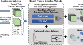
High sensitivity methods for automated rib fracture detection in pediatric radiographs
- Jonathan Burkow
- Gregory Holste
- Adam Alessio
News and Comment
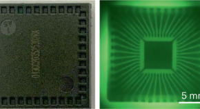
Scintillating observations
An organic scintillator with high imaging resolution and a low detection limit is fabricated by using heavy atoms to increase the X-ray absorption of thermally activated delayed fluorescence chromophores.
- Hannah Hatcher
Prasugrel superior to ticagrelor in ACS
- Irene Fernández-Ruiz
Cardiovascular MRI versus FFR in stable angina
- Gregory B. Lim
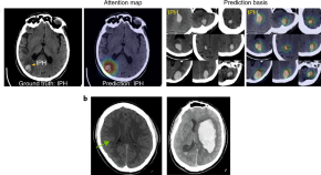
Spotting brain bleeding after sparse training
Accurate and explainable detection, via deep learning, of acute intracranial haemorrhage from computed tomography images of the head is achievable with small amounts of data for model training.
- Michael C. Muelly
Early invasive assessment of NSTE-ACS
Peerfect scores for rcc.
- Louise Stone
Quick links
- Explore articles by subject
- Guide to authors
- Editorial policies
Retrieval Augmented Chest X-Ray Report Generation using OpenAI GPT models
- Mercy Ranjit ,
- Gopinath Ganapathy ,
- Ranjit Manuel ,
- Tanuja Ganu
Publication
We propose Retrieval Augmented Generation (RAG) as an approach for automated radiology report writing that leverages multimodally aligned embeddings from a contrastively pretrained vision language model for retrieval of relevant candidate radiology text for an input radiology image and a general domain generative model like OpenAI text-davinci-003, gpt-3.5-turbo and gpt-4 for report generation using the relevant radiology text retrieved. This approach keeps hallucinated generations under check and provides capabilities to generate report content in the format we desire leveraging the instruction following capabilities of these generative models. Our approach achieves better clinical metrics with a BERTScore of 0.2865 ({\Delta}+ 25.88%) and Semb score of 0.4026 ({\Delta}+ 6.31%). Our approach can be broadly relevant for different clinical settings as it allows to augment the automated radiology report generation process with content relevant for that setting while also having the ability to inject user intents and requirements in the prompts as part of the report generation process to modulate the content and format of the generated reports as applicable for that clinical setting.
- Follow on Twitter
- Like on Facebook
- Follow on LinkedIn
- Subscribe on Youtube
- Follow on Instagram
- Subscribe to our RSS feed
Share this page:
- Share on Twitter
- Share on Facebook
- Share on LinkedIn
- Share on Reddit
The browser you are using is no longer supported and for that reason you will not get the best experience when using our website.
You currently have JavaScript disabled in your web browser, please enable JavaScript to view our website as intended.
Pint of Science Leicester brings the stars behind the latest research to a venue near you
20 scientists from the University of Leicester take to the stage in venues across Leicester next week to bring science to the public – and the pub!
On 13–15 May, the Pint of Science festival will simultaneously bring thousands of scientists and their research out of the lab and into pubs, cafes and community halls near you. The three-day festival has grown astronomically since its inception, and 2024 will see events in over 25 countries and hundreds of cities around the world.
Each night will provide a unique line up of talks, demonstrations and live experiments held in a relaxed and informal environment.
University of Leicester researchers will be speaking at venues across the city, including Phoenix Theatre, The Real Ale Classroom, and Rainbow and Dove. Tickets are available from the Pint of Science website , costing just £5 each. Attendees in Leicester will enjoy a variety of exciting talks including: rural racism, climate refugees, and the latest developments in cancer research.
The full list of speakers and events by University of Leicester researchers is available at the Leicester Pint of Science website .
The organisers of this year’s University of Leicester talks said: "We are excited and proud to host four different themes in the 2024 edition of Pint of Science! We are looking forward to getting our speakers into three beautiful venues with members of the public and talk about cutting-edge research led by University of Leicester researchers. This year's programme will feature AI-powered robots, latest developments in cancer research, rural racism, and so much more."
Pint of Science founders Dr Praveen Paul and Dr Michael Motskin wanted to bring back the personal touch to science. From three cities back in 2013 to hundreds of cities this year, the festival continues to showcase the brilliant work happening on our doorsteps. Events provide a platform for researchers to share their stories, and audiences an opportunity to ask their questions about science to those directly behind it.
“It’s fantastic to see how a simple idea between a few friends has captured people’s imaginations and pushed scientific research into the spotlight”, says Co-Founder Dr Praveen Paul. “We should all have an opportunity to question and discover the breadth of research that is taking place right here in the UK. None of it would be possible without our enthusiastic and dedicated volunteers who work so hard to bring you a programme packed full of inspiring events, and encourage us all to be curious. The only difficulty is choosing which of the brilliant events to go to!”
- For more information, contact Dr Marta Mangiarulo at [email protected]
- Community engagement
- Public events
Related stories
University of leicester to spend £1m on making research more inclusive, students take on the challenges of living on the moon, innovative leicester centre spearheads international initiative, common childhood eye disorder is best treated by patching earlier, new study shows, leicester-tested einstein probe opens its wide eyes to the x-ray sky, researchers use nasa’s webb to map weather of planet 280 light-years away.

IMAGES
VIDEO
COMMENTS
X-ray imaging is a low-cost, powerful technology that has been extensively used in medical diagnosis and industrial nondestructive inspection. The ability of X-rays to penetrate through the body presents great advances for noninvasive imaging of its internal structure. ... Another focus of recent research is developing flexible X-ray detectors ...
X-rays are a type of electromagnetic radiation with a wavelength between 0.01 and 10 nanometres. X-rays offer an important method for investigating the atomic structure of crystalline materials ...
X-ray (or much less commonly, X-radiation) is a high-energy electromagnetic radiation. ... Edison dropped X-ray research around 1903, before the death of Clarence Madison Dally, one of his glassblowers. Dally had a habit of testing X-ray tubes on his own hands, ...
X-ray-induced explosions in water drops, examined using time-resolved imaging, show interacting high-speed liquid and vapour flows. This type of X-ray absorption dynamics is predictable and may be ...
X-ray computed tomography (CT) can reveal the internal details of objects in three dimensions non-destructively. ... Recent research has also documented the variability introduced by different ...
X-Rays June 2022 What are medical x-rays? X-rays are a form of electromagnetic radiation, similar t o visible light. Unlike light, however, x-rays have higher energy ... Current research of x -ray technology focuses on ways to reduce radiation dose, improve image resolution, and enhance contrast materials and methods. The following are examples of
X-rays are a form of electromagnetic radiation, similar to visible light. Unlike light, however, x-rays have higher energy and can pass through most objects, including the body. Medical x-rays are used to generate images of tissues and structures inside the body. If x-rays travelling through the body also pass through an x-ray detector on the ...
Scintillators are materials that emit light when bombarded with high-energy particles or X-rays. In medical or dental X-ray systems, they convert incoming X-ray radiation into visible light that can then be captured using film or photosensors. They're also used for night-vision systems and for research, such as in particle detectors or ...
X-ray, electromagnetic radiation of extremely short wavelength and high frequency, with wavelengths ranging from about 10^-8 to 10^-12 metre. The passage of X-rays through materials, including biological tissue, can be recorded. Thus, analysis of X-ray images of the body is a valuable medical diagnostic tool.
An X-ray is a quick, painless test that captures images of the structures inside the body — particularly the bones. X-ray beams pass through the body. These beams are absorbed in different amounts depending on the density of the material they pass through. Dense materials, such as bone and metal, show up as white on X-rays.
The newly upgraded Linac Coherent Light Source (LCLS) X-ray free-electron laser (XFEL) at the Department of Energy's SLAC National Accelerator Laboratory successfully produced its first X-rays, and researchers around the world are already lined up to kick off an ambitious science program.. The upgrade, called LCLS-II, creates unparalleled capabilities that will usher in a new era in research ...
Biomedical Research. X-ray diffraction is the technique where X-ray light changes its direction by amounts that depend on the X-ray energy, much like a prism separates light into its component colors. Scientists using Chandra take advantage of diffraction to reveal important information about distant cosmic sources using the observatory's two ...
Get an overview of research at SLAC: X-ray and ultrafast science, particle and astrophysics, cosmology, particle accelerators, biology, energy and technology. X-ray & ultrafast science. Revealing nature's fastest processes with X-rays, lasers and electrons. Physics of the universe. Studying the particles and forces that knit the cosmos together
DISCOVERY OF X-RAYS. X-rays were first observed and documented in 1895 by German scientist Wilhelm Conrad Roentgen. He discovered that firing streams of x-rays through arms and hands created detailed images of the bones inside. When you get an x-ray taken, x-ray sensitive film is put on one side of your body, and x-rays are shot through you.
We further discuss future research directions and challenges in developing advanced next-generation materials that are crucial to the fabrication of flexible, low-dose, high-resolution X-ray imaging detectors. 1. Introduction. X-rays are a type of ionizing radiation with a wavelength ranging from 0.01 to 10 nm [ 1, 2 ].
X-rays are a form of electromagnetic radiation with wavelengths ranging from 0.01 to 10 nanometers. In the setting of diagnostic radiology, X-rays have long enjoyed use in the imaging of body tissues and aid in the diagnosis of disease. Simply understood, the generation of X-rays occurs when electrons are accelerated under a potential difference and turned into electromagnetic radiation.[1] An ...
X-ray tomography, or X-ray computed tomography, is a method for generating 3-dimensional imaged volumes from 2-dimensional X-ray image slices. X-ray imaging is based on the differential absorption ...
While X-rays are linked to a slightly increased risk of cancer, there is an extremely low risk of short-term side effects. Exposure to high radiation levels can have a range of effects, such as ...
Learn about X-ray imaging, used to photograph internal structures of the body. While common and useful, this test does carry some some risks. ... Similarly, if you are paying for your X-ray and have time to research the fees, you can call the hospital's billing department ahead of time to get a quote for the procedure. Doing so can help you ...
An X-ray is a test that uses small doses of radiation to take pictures of the inside of your body. Find out how you have them and what happens afterwards. ... Cancer Research UK is a registered charity in England and Wales (1089464), Scotland (SC041666), the Isle of Man (1103) and Jersey (247). A company limited by guarantee.
An X-ray tube (SC-T160/1.2-80, Beijing Research Institute of Mechanical & Electrical Technology) was used to generate the radiation field. The maximum tube voltage is 160 kV, and the maximum tube current is 1.2 mA. ... X-rays with high energy and strong penetrating ability can realize long distance propagation in the air, using this ...
The space X-ray telescope zoomed in on a few well-known celestial objects to give us a hint of what the mission is capable of. ... Medical research advances and health news. Tech Xplore.
After starting science operations in February, Japan-led XRISM (X-ray Imaging and Spectroscopy Mission) studied the monster black hole at the center of galaxy NGC 4151.
When Bulbul learned ORCs hadn't been studied in X-ray light, she and postdoctoral researcher Xiaoyuan Zhang began poring over data from eROSITA (Extended Roentgen Survey with an Imaging Telescope Array), an orbiting German/Russian X-ray telescope. They noticed some X-ray emission that seemed like it could be from the Cloverleaf, based on less ...
This experimental study established a novel 3D imaging method using correlative X-Ray and scanning electron microscopy to characterize these damages. By integrating the information obtained from both X-ray microscopy and SEM, a comprehensive picture of the materials structure and performance could be established.
Radiography is the use of X-rays or gamma rays to examine non-uniformly composed material; images are recorded on a sensitized surface, such as photographic film or a digital detector ...
We propose Retrieval Augmented Generation (RAG) as an approach for automated radiology report writing that leverages multimodally aligned embeddings from a contrastively pretrained vision language model for retrieval of relevant candidate radiology text for an input radiology image and a general domain generative model like OpenAI text-davinci-003, gpt-3.5-turbo and gpt-4 for report generation ...
The local structure of β-(Al x Ga 1-x) 2 O 3 alloys for various Al contents has been determined from extended X-ray absorption fine structure spectroscopy (EXAFS) performed at the Ga K-edge. A model to fit the experimental EXAFS data of the monoclinic β-(Al x Ga 1-x) 2 O 3 alloy has been suggested to independently determine relevant local ...
On 13-15 May, the Pint of Science festival will simultaneously bring thousands of scientists and their research out of the lab and into pubs, cafes and community halls near you. The three-day festival has grown astronomically since its inception, and 2024 will see events in over 25 countries and hundreds of cities around the world ...